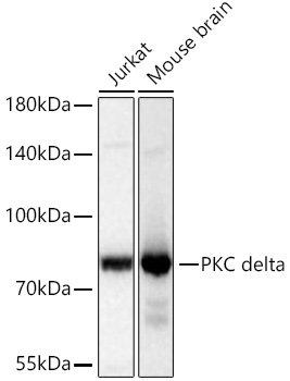
Western blot analysis of various lysates using PKC delta Rabbit mAb (A7778) at 1:1000 dilution incubated at room temperature for 1.5 hours.
Secondary antibody: HRP-conjugated Goat anti-Rabbit IgG (H+L) (AS014) at 1:10000 dilution.
Lysates/proteins: 25 μg per lane.
Blocking buffer: 3% nonfat dry milk in TBST.
Detection: ECL Basic Kit (RM00020).
Exposure time: 5 s.
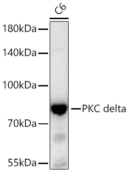
Western blot analysis of lysates from C6 cells using PKC delta Rabbit mAb (A7778) at 1:1000 dilution incubated at room temperature for 1.5 hours.
Secondary antibody: HRP-conjugated Goat anti-Rabbit IgG (H+L) (AS014) at 1:10000 dilution.
Lysates/proteins: 25 μg per lane.
Blocking buffer: 3% nonfat dry milk in TBST.
Detection: ECL Basic Kit (RM00020).
Exposure time: 45 s.
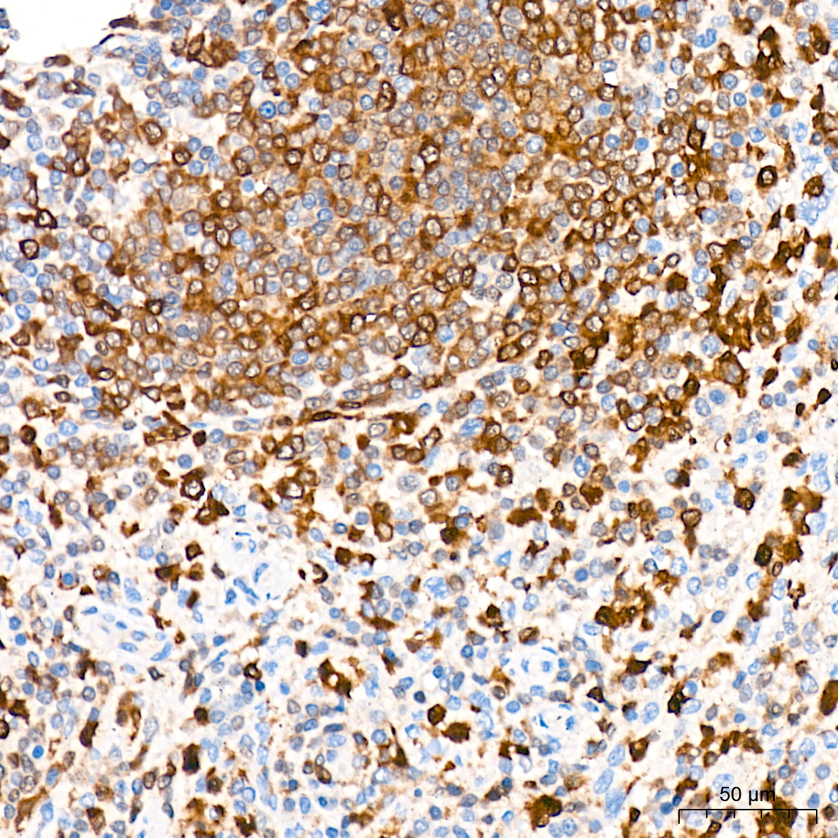
Immunohistochemistry analysis of paraffin-embedded Human spleen tissue using PKC delta Rabbit mAb (A7778) at a dilution of 1:200 (40x lens). High pressure antigen retrieval was performed with 0.01 M citrate buffer (pH 6.0) prior to IHC staining.
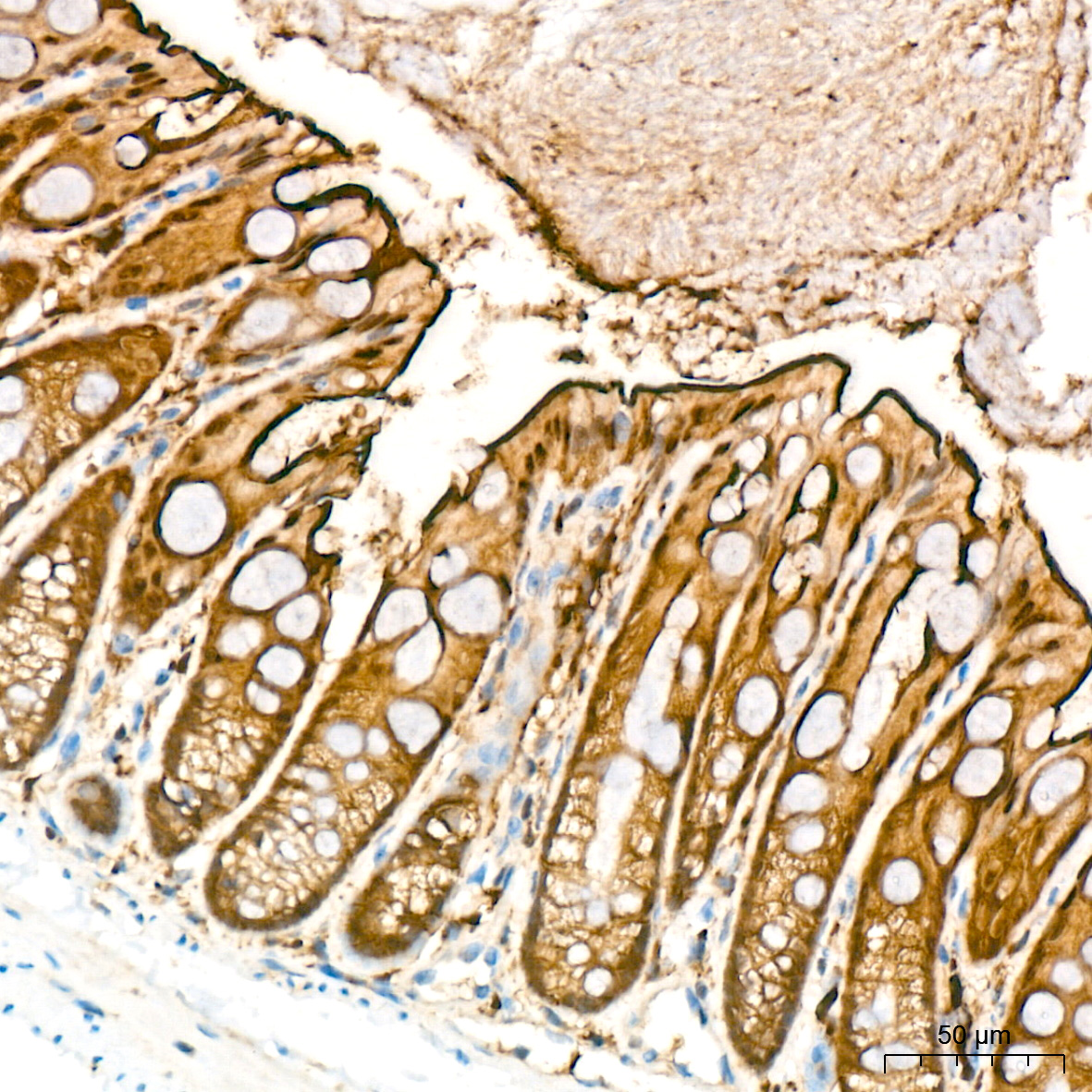
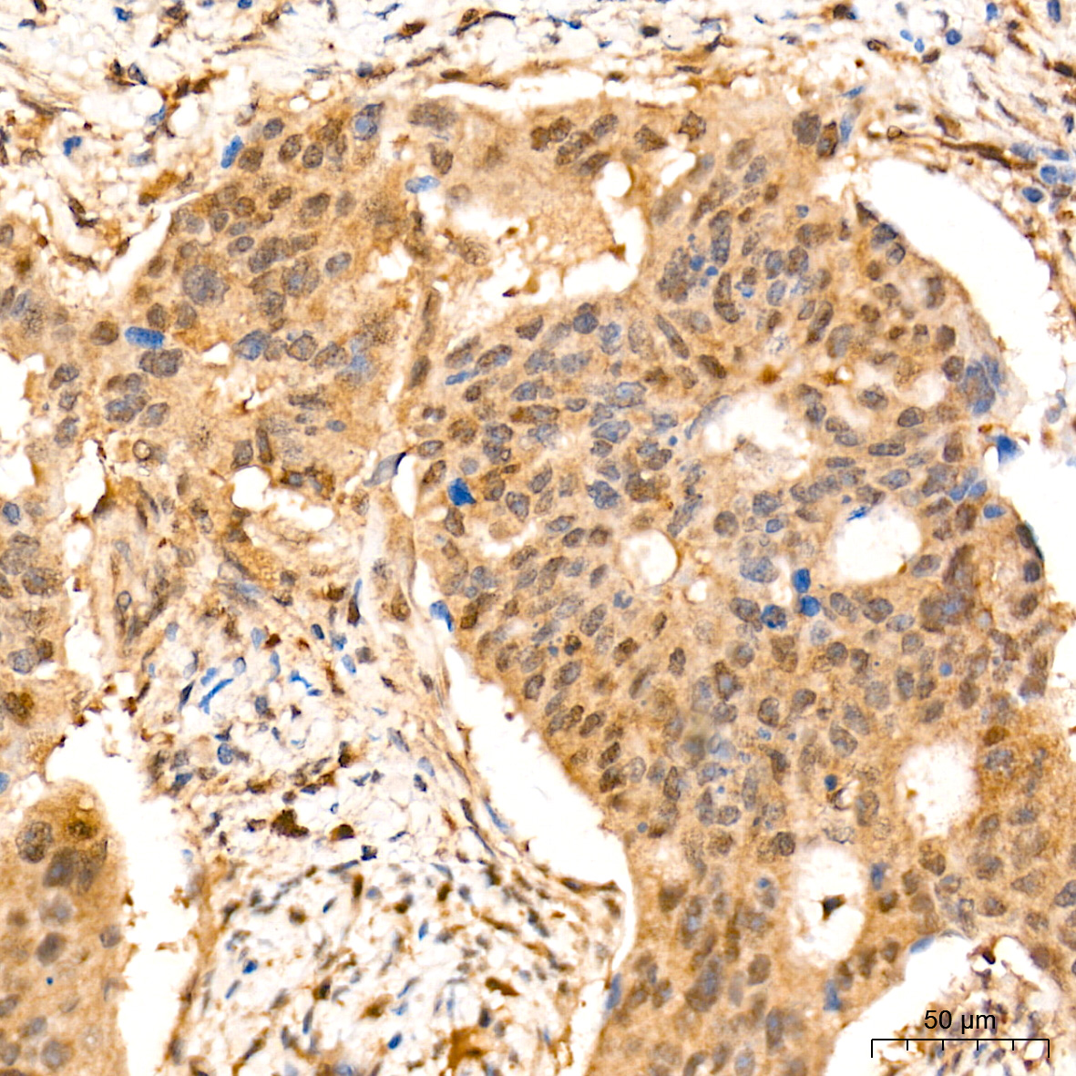
Immunohistochemistry analysis of paraffin-embedded Mouse colon tissue using PKC delta Rabbit mAb (A7778) at a dilution of 1:200 (40x lens). High pressure antigen retrieval was performed with 0.01 M citrate buffer (pH 6.0) prior to IHC staining.
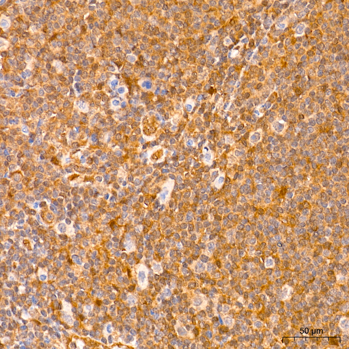
Immunohistochemistry analysis of paraffin-embedded Human thyroid tissue using PKC delta Rabbit mAb (A7778) at a dilution of 1:200 (40x lens). High pressure antigen retrieval was performed with 0.01 M citrate buffer (pH 6.0) prior to IHC staining.
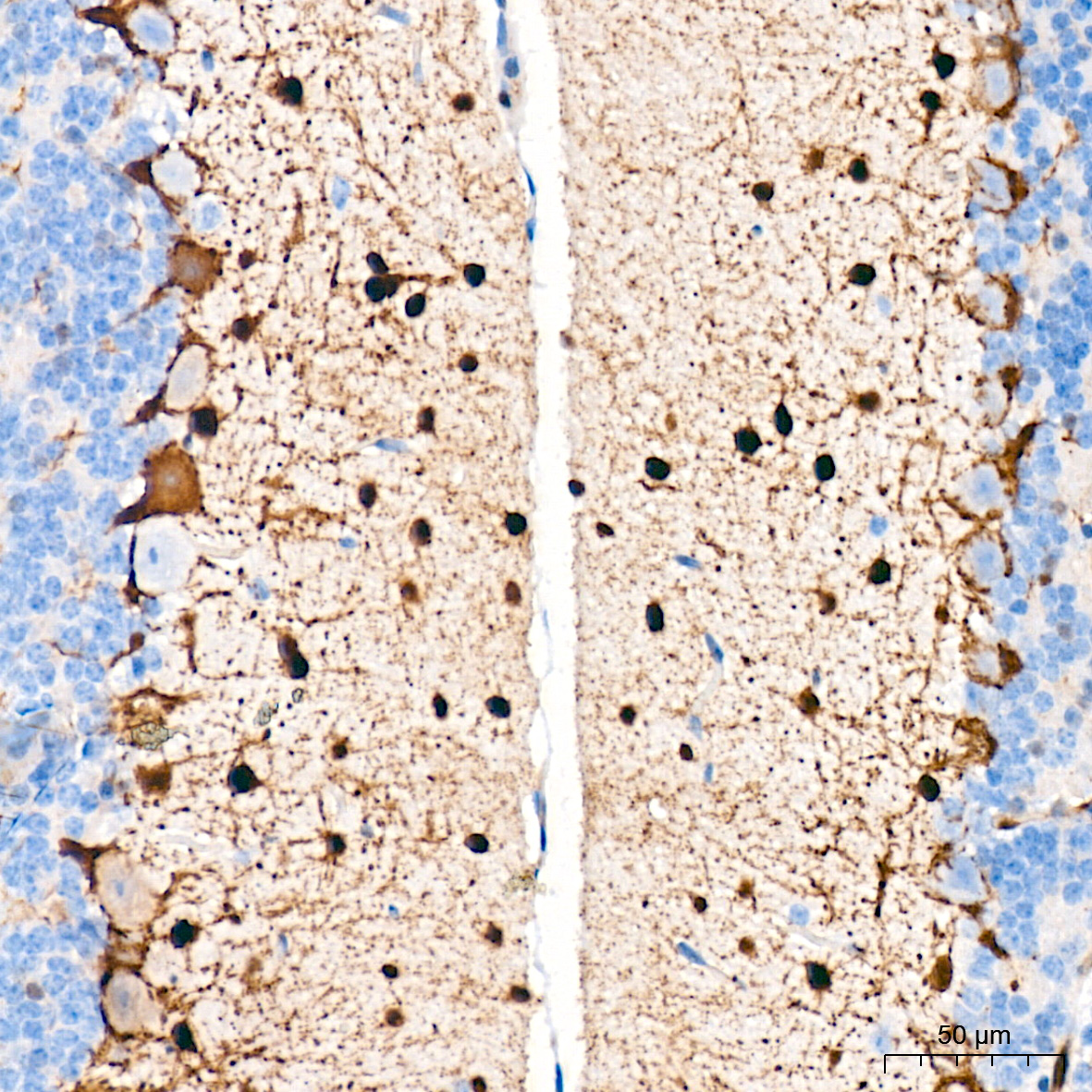
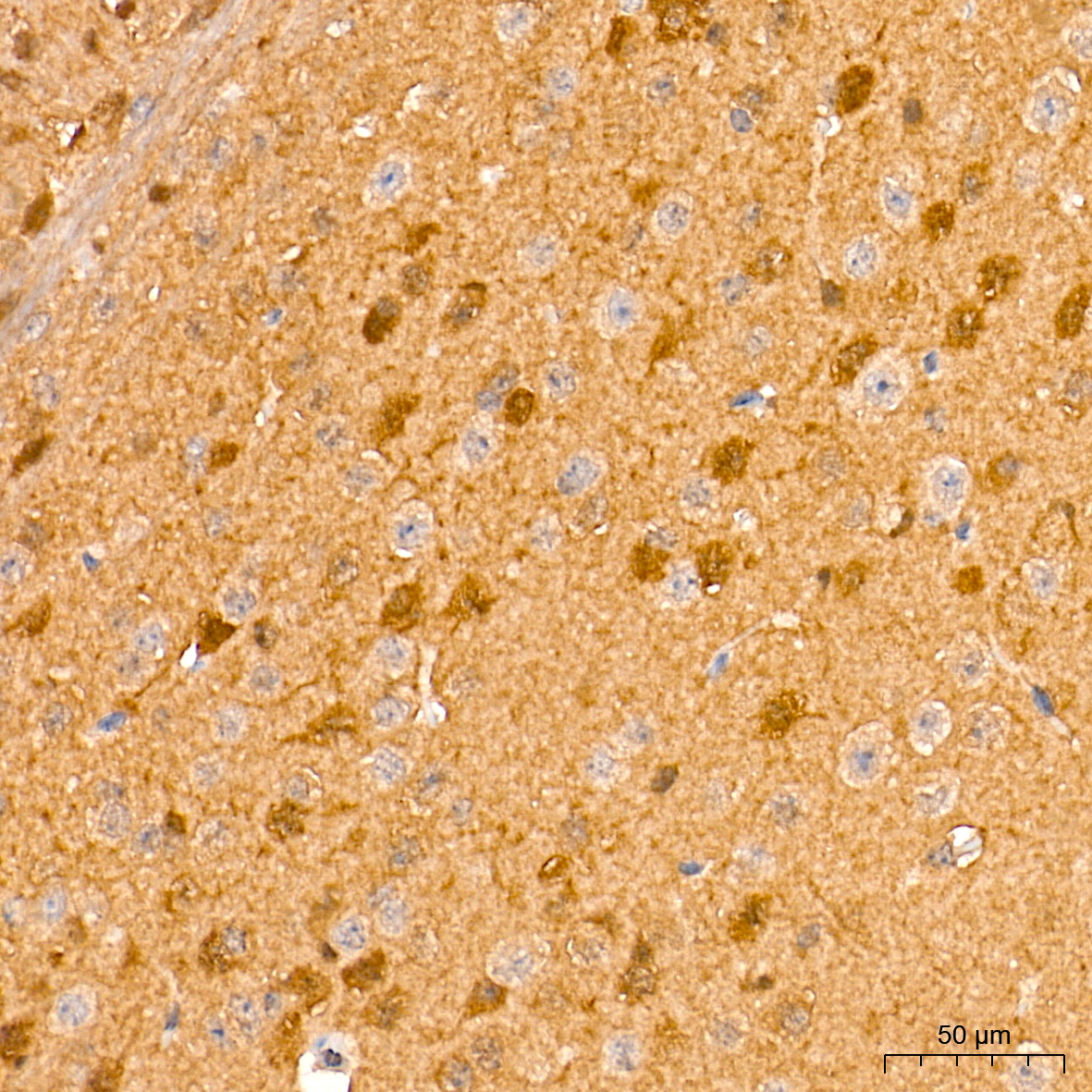
Immunohistochemistry analysis of paraffin-embedded Mouse brain tissue using PKC delta Rabbit mAb (A7778) at a dilution of 1:200 (40x lens). High pressure antigen retrieval was performed with 0.01 M citrate buffer (pH 6.0) prior to IHC staining.
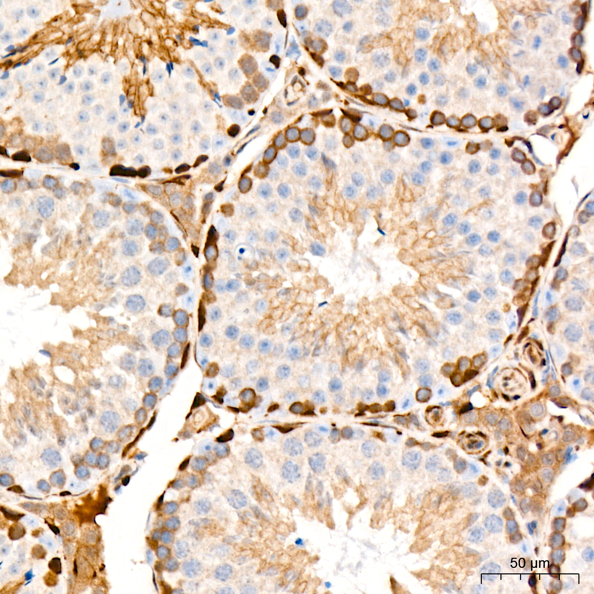
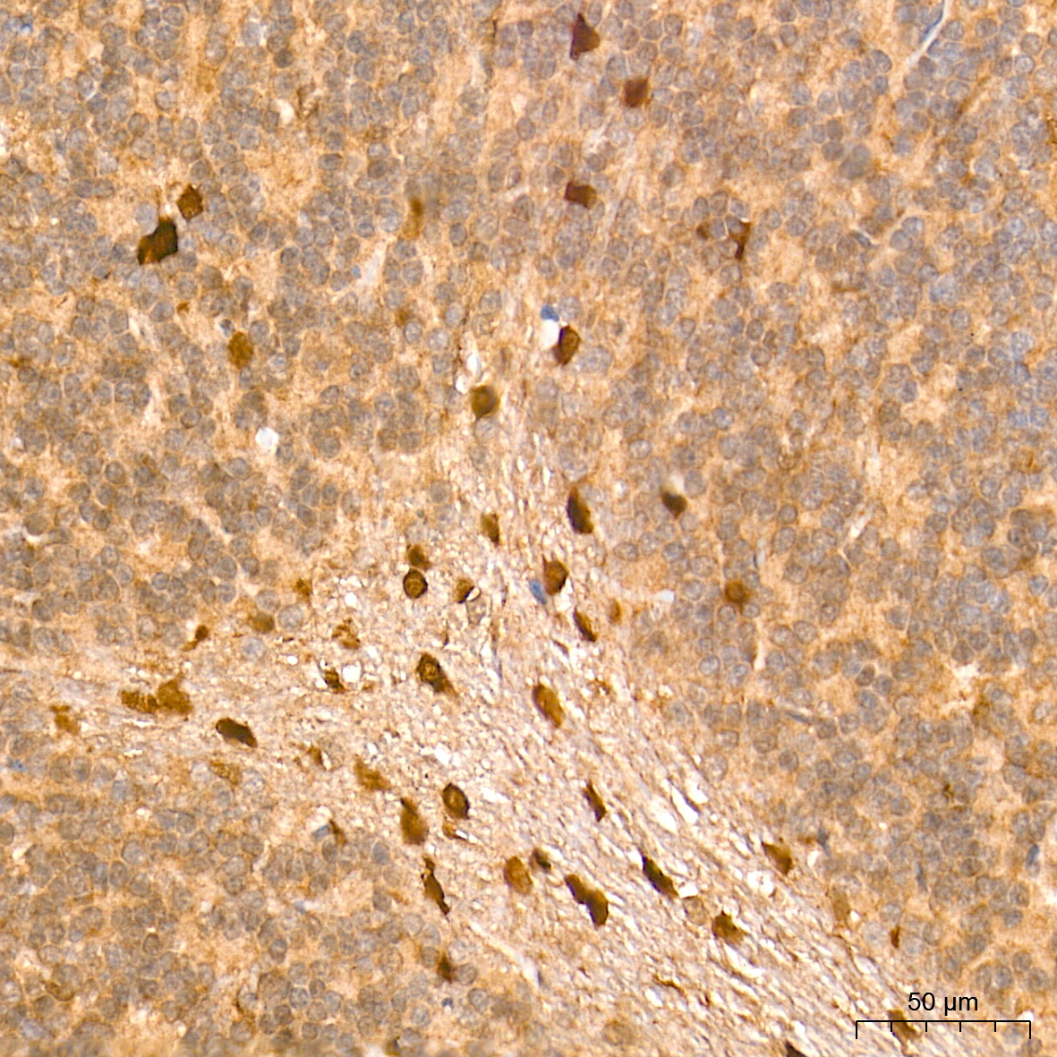
Immunohistochemistry analysis of paraffin-embedded Mouse testis tissue using PKC delta Rabbit mAb (A7778) at a dilution of 1:200 (40x lens). High pressure antigen retrieval was performed with 0.01 M citrate buffer (pH 6.0) prior to IHC staining.
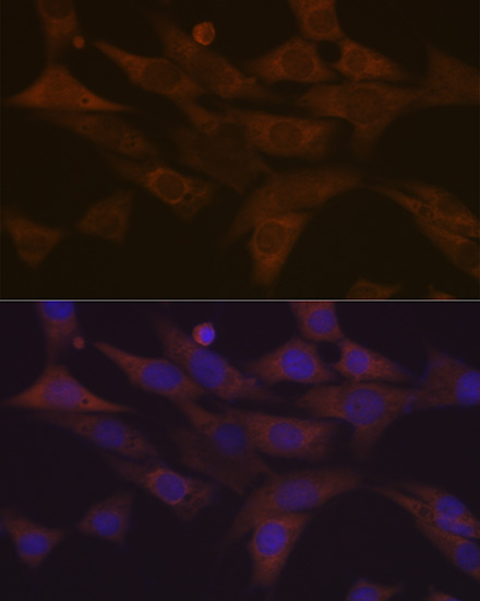
Immunofluorescence analysis of NIH-3T3 cells using PKC delta Rabbit mAb (A7778) at dilution of 1:100 (40x lens). Secondary antibody: Cy3-conjugated Goat anti-Rabbit IgG (H+L) (AS007) at 1:500 dilution. Blue: DAPI for nuclear staining.
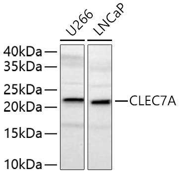
Western blot analysis of various lysates using CLEC7A Rabbit pAb (A9883) at 1:2000 dilution incubated overnight at 4℃.
Secondary antibody: HRP-conjugated Goat anti-Rabbit IgG (H+L) (AS014) at 1:10000 dilution.
Lysates/proteins: 25 μg per lane.
Blocking buffer: 3% nonfat dry milk in TBST.
Detection: ECL Basic Kit (RM00020).
Exposure time: 90 s.
Phospho-PLC gamma 1 (PLCG1)-S1248 Rabbit pAb
_WB_01.jpg?t=1767195090)
Western blot analysis of lysates from Jurkat cells, using Phospho-PLC gamma 1 (PLCG1)-S1248 Rabbit pAb (AP0827) at dilution or PLC gamma 1 (PLCG1) antibody (A15704). Jurkat cells were treated with PMA/TPA (200 nM) at 37℃ for 10 minutes.
Secondary antibody: HRP-conjugated Goat anti-Rabbit IgG (H+L) (AS014) at 1:10000 dilution.
Lysates/proteins: 25μg per lane.
Blocking buffer: 3% BSA.
Detection: ECL Basic Kit (RM00020).
Exposure time: 90s.
_IHC_01.jpg?t=1767195090)
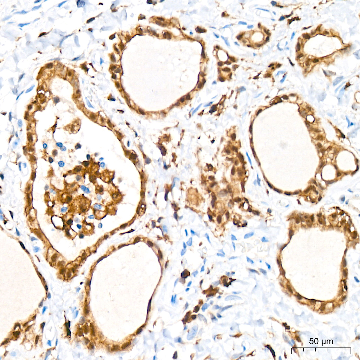
Immunohistochemistry analysis of paraffin-embedded Human placenta using Phospho-PLC gamma 1 (PLCG1)-S1248 Rabbit pAb (AP0827) at dilution of 1:100 (40x lens). Microwave antigen retrieval performed with 0.01M Tris/EDTA Buffer (pH 9.0) prior to IHC staining.
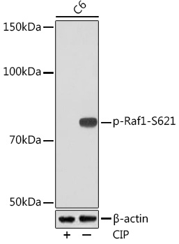
Western blot analysis of lysates from C6 cells, using Phospho-Raf1-S621 Rabbit mAb (AP1011) at 1:1000 dilution. C6 cells were treated by CIP(20uL/400ul) at 37℃ for 1 hour.
Secondary antibody: HRP-conjugated Goat anti-Rabbit IgG (H+L) (AS014) at 1:10000 dilution.
Lysates/proteins: 25μg per lane.
Blocking buffer: 3% BSA.
Detection: ECL Basic Kit (RM00020).
Exposure time: 3min.
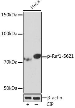
Western blot analysis of lysates from HeLa cells, using Phospho-Raf1-S621 Rabbit mAb (AP1011) at 1:1000 dilution. HeLa cells were treated by CIP(20uL/400ul) at 37℃ for 1 hour.
Secondary antibody: HRP-conjugated Goat anti-Rabbit IgG (H+L) (AS014) at 1:10000 dilution.
Lysates/proteins: 25μg per lane.
Blocking buffer: 3% BSA.
Detection: ECL Enhanced Kit (RM00021).
Exposure time: 3min.
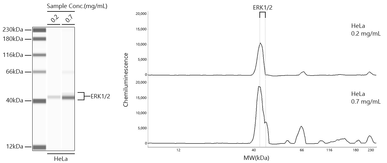
Simple Western™ analysis of lysates from HeLa cells using ERK1/2 Rabbit mAb (A4782) at 1:100 dilution. The virtual lane view (left) shows the target band (as indicated) with samples in concentrations of 0.2 mg/mL and 0.7 mg/mL. The corresponding electropherogram view (right) plots chemiluminescence intensity against molecular weight along the capillary for sample concentrations of 0.2 mg/mL and 0.7 mg/mL. This experiment was performed under reducing conditions on the Jess™ Simple Western instrument from ProteinSimple, a BioTechne brand, using the 12-230 kDa separation module.
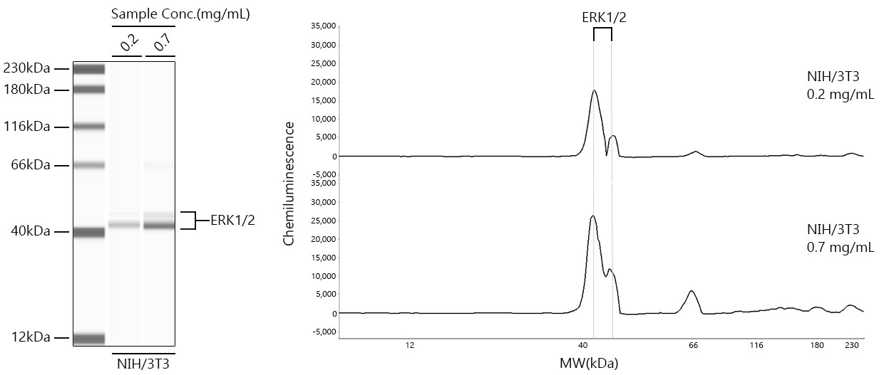
Simple Western™ analysis of lysates from NIH/3T3 cells using ERK1/2 Rabbit mAb (A4782) at 1:100 dilution. The virtual lane view (left) shows the target band (as indicated) with samples in concentrations of 0.2 mg/mL and 0.7 mg/mL. The corresponding electropherogram view (right) plots chemiluminescence intensity against molecular weight along the capillary for sample concentrations of 0.2 mg/mL and 0.7 mg/mL. This experiment was performed under reducing conditions on the Jess™ Simple Western instrument from ProteinSimple, a BioTechne brand, using the 12-230 kDa separation module.
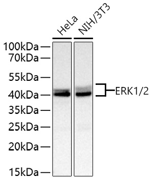
Western blot analysis of various lysates using ERK1/2 Rabbit mAb (A4782) at 1:4000 dilution incubated overnight at 4℃.
Secondary antibody: HRP-conjugated Goat anti-Rabbit IgG (H+L) (AS014) at 1:10000 dilution.
Lysates/proteins: 25 μg per lane.
Blocking buffer: 3% nonfat dry milk in TBST.
Detection: ECL Basic Kit (RM00020).
Exposure time: 1 s.
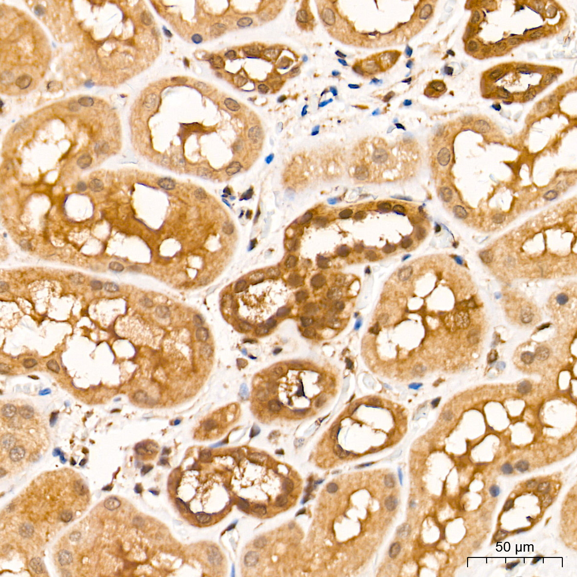
Immunohistochemistry analysis of paraffin-embedded Human kidney tissue using ERK1/2 Rabbit mAb (A4782) at a dilution of 1:9600 (40x lens). High pressure antigen retrieval performed with 0.01M Tris-EDTA Buffer (pH 9.0) prior to IHC staining.
Immunohistochemistry analysis of paraffin-embedded Human colon carcinoma tissue using ERK1/2 Rabbit mAb (A4782) at a dilution of 1:9600 (40x lens). High pressure antigen retrieval performed with 0.01M Tris-EDTA Buffer (pH 9.0) prior to IHC staining.
Immunohistochemistry analysis of paraffin-embedded Human tonsil tissue using ERK1/2 Rabbit mAb (A4782) at a dilution of 1:9600 (40x lens). High pressure antigen retrieval performed with 0.01M Tris-EDTA Buffer (pH 9.0) prior to IHC staining.
Immunohistochemistry analysis of paraffin-embedded Mouse brain tissue using ERK1/2 Rabbit mAb (A4782) at a dilution of 1:9600 (40x lens). High pressure antigen retrieval performed with 0.01M Tris-EDTA Buffer (pH 9.0) prior to IHC staining.
Immunohistochemistry analysis of paraffin-embedded Rat brain tissue using ERK1/2 Rabbit mAb (A4782) at a dilution of 1:9600 (40x lens). High pressure antigen retrieval performed with 0.01M Tris-EDTA Buffer (pH 9.0) prior to IHC staining.
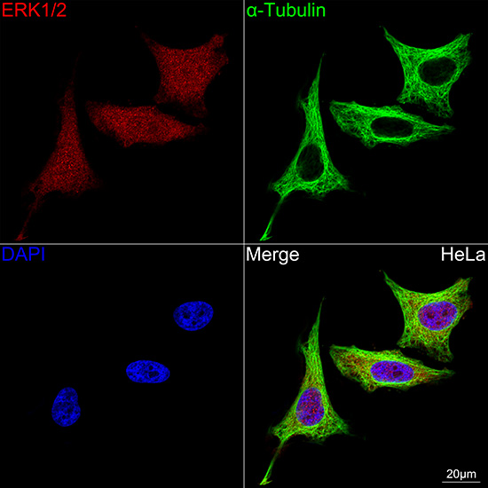
Confocal imaging of HeLa cells using ERK1/2 Rabbit mAb (A4782, dilution 1:200) followed by a further incubation with Cy3-conjugated Goat Anti-Rabbit IgG (H+L) (AS007, dilution 1:500) (Red). The cells were counterstained with α-Tubulin Mouse mAb (AC012, dilution 1:400) followed by incubation with ABflo® 488-conjugated Goat Anti-Mouse IgG (H+L) (AS076, dilution 1:500) (Green). DAPI was used for nuclear staining (Blue). Objective: 100x.
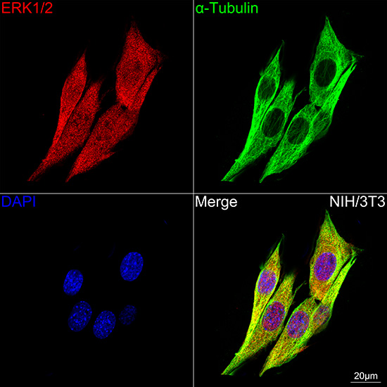
Confocal imaging of NIH/3T3 cells using ERK1/2 Rabbit mAb (A4782, dilution 1:200) followed by a further incubation with Cy3-conjugated Goat Anti-Rabbit IgG (H+L) (AS007, dilution 1:500) (Red). The cells were counterstained with α-Tubulin Mouse mAb (AC012, dilution 1:400) followed by incubation with ABflo® 488-conjugated Goat Anti-Mouse IgG (H+L) (AS076, dilution 1:500) (Green). DAPI was used for nuclear staining (Blue). Objective: 100x.
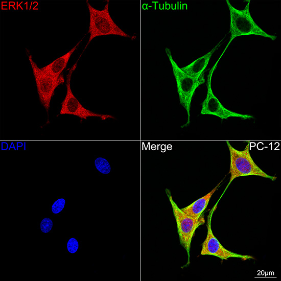
Confocal imaging of PC-12 cells using ERK1/2 Rabbit mAb (A4782, dilution 1:200) followed by a further incubation with Cy3-conjugated Goat Anti-Rabbit IgG (H+L) (AS007, dilution 1:500) (Red). The cells were counterstained with α-Tubulin Mouse mAb (AC012, dilution 1:400) followed by incubation with ABflo® 488-conjugated Goat Anti-Mouse IgG (H+L) (AS076, dilution 1:500) (Green). DAPI was used for nuclear staining (Blue). Objective: 100x.
Phospho-ERK1-T202 + ERK2-T185 Rabbit mAb
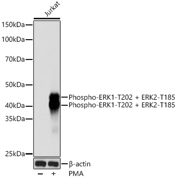
Western blot analysis of lysates from Jurkat cells, using Phospho-ERK1-T202 + ERK2-T185 Rabbit mAb (AP0485) at 1:1000 dilution. Jurkat cells were treated with PMA/TPA (200 nM) at 37℃ for 10 minutes.
Secondary antibody: HRP-conjugated Goat anti-Rabbit IgG (H+L) (AS014) at 1:10000 dilution.
Lysates/proteins: 25μg per lane.
Blocking buffer: 3% nonfat dry milk in TBST.
Detection: ECL Basic Kit (RM00020).
Exposure time: 10s.
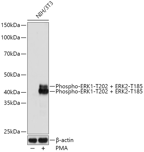
Western blot analysis of lysates from NIH/3T3 cells, using Phospho-ERK1-T202 + ERK2-T185 Rabbit mAb (AP0485) at 1:1000 dilution. NIH/3T3 cells were treated with PMA/TPA (200 nM) at 37℃ for 30 minutes after serum-starvation overnight.
Secondary antibody: HRP-conjugated Goat anti-Rabbit IgG (H+L) (AS014) at 1:10000 dilution.
Lysates/proteins: 25μg per lane.
Blocking buffer: 3% nonfat dry milk in TBST.
Detection: ECL Basic Kit (RM00020).
Exposure time: 1S.
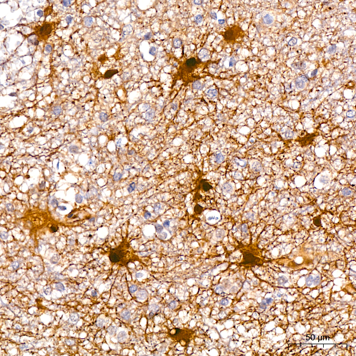
Immunohistochemistry analysis of paraffin-embedded Human brain using Phospho-ERK1-T202 + ERK2-T185 Rabbit mAb (AP0485) at dilution of 1:200 (40x lens). High pressure antigen retrieval performed with 0.01M Citrate buffer (pH 6.0) prior to IHC staining.
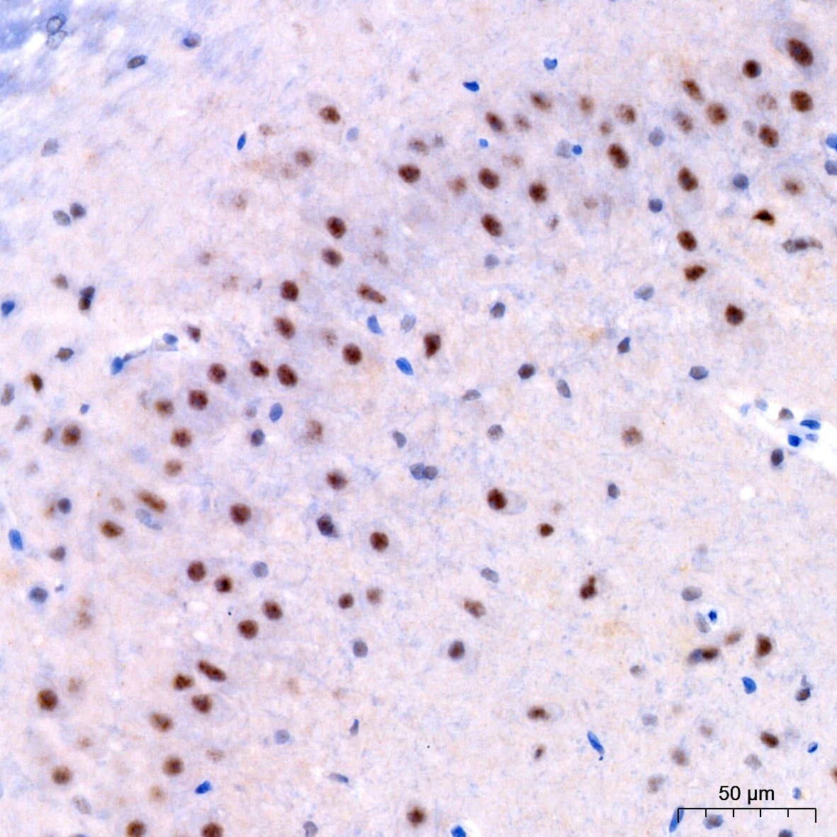
Immunohistochemistry analysis of paraffin-embedded Rat brain using Phospho-ERK1-T202 + ERK2-T185 Rabbit mAb (AP0485) at dilution of 1:200 (40x lens). High pressure antigen retrieval performed with 0.01M Citrate buffer (pH 6.0) prior to IHC staining.