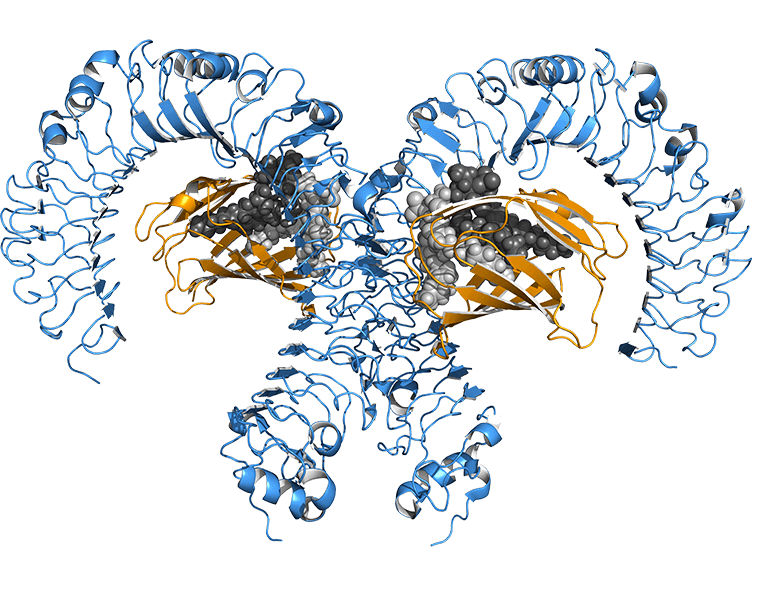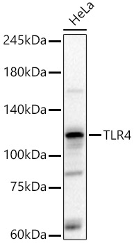
Western blot analysis of various lysates, using TLR4 Rabbit pAb (A5258) at 1:2000 dilution.
Secondary antibody: HRP-conjugated Goat anti-Rabbit IgG (H+L) (AS014) at 1:10000 dilution.
Lysates/proteins: 25μg per lane.
Blocking buffer: 3% nonfat dry milk in TBST.
Detection: ECL Basic Kit (RM00020).
Exposure time: 60s.
_WB_01.jpg?t=1779906405)
Western blot analysis of lysates from Mouse liver, using TLR4 Rabbit pAb (A5258) at 1:700 dilution.
Secondary antibody: HRP-conjugated Goat anti-Rabbit IgG (H+L) (AS014) at 1:10000 dilution.
Lysates/proteins: 25μg per lane.
Blocking buffer: 3% nonfat dry milk in TBST.
Detection: ECL Enhanced Kit (RM00021).
Exposure time: 60s.
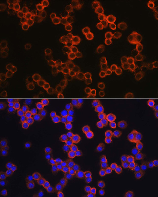
Immunofluorescence analysis of RAW264.7 cells using TLR4 Rabbit pAb (A5258) at dilution of 1:200 (40x lens).Secondary antibody: Cy3-conjugated Goat anti-Rabbit IgG (H+L) (AS007) at 1:500 dilution. Blue: DAPI for nuclear staining.
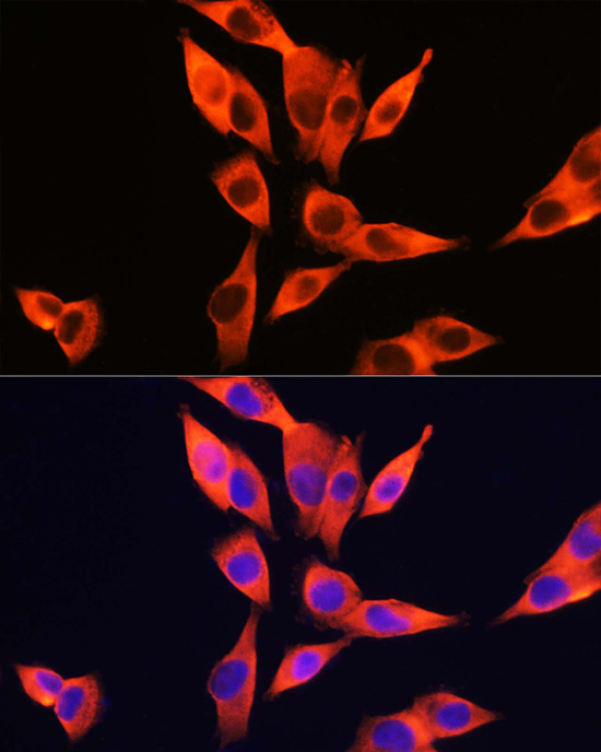
Immunofluorescence analysis of HepG2 cells using TLR4 Rabbit pAb (A5258) at dilution of 1:200 (40x lens).Secondary antibody: Cy3-conjugated Goat anti-Rabbit IgG (H+L) (AS007) at 1:500 dilution. Blue: DAPI for nuclear staining.
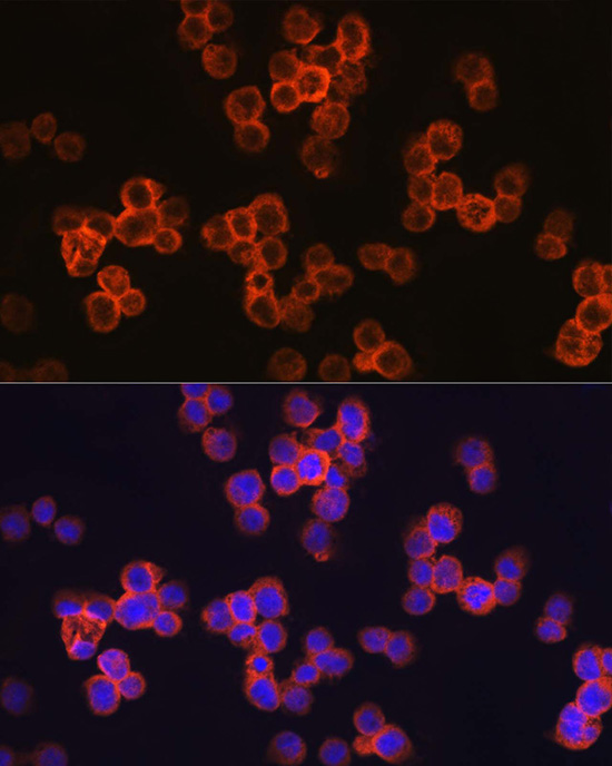
Immunofluorescence analysis of THP-1 cells using TLR4 Rabbit pAb (A5258) at dilution of 1:200 (40x lens).Secondary antibody: Cy3-conjugated Goat anti-Rabbit IgG (H+L) (AS007) at 1:500 dilution. Blue: DAPI for nuclear staining.
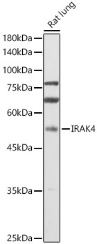
Western blot analysis of lysates from Rat lung, using IRAK4 Rabbit pAb (A6208) at 1:1000 dilution.
Secondary antibody: HRP-conjugated Goat anti-Rabbit IgG (H+L) (AS014) at 1:10000 dilution.
Lysates/proteins: 25μg per lane.
Blocking buffer: 3% nonfat dry milk in TBST.
Detection: ECL Enhanced Kit (RM00021).
Exposure time: 60s.
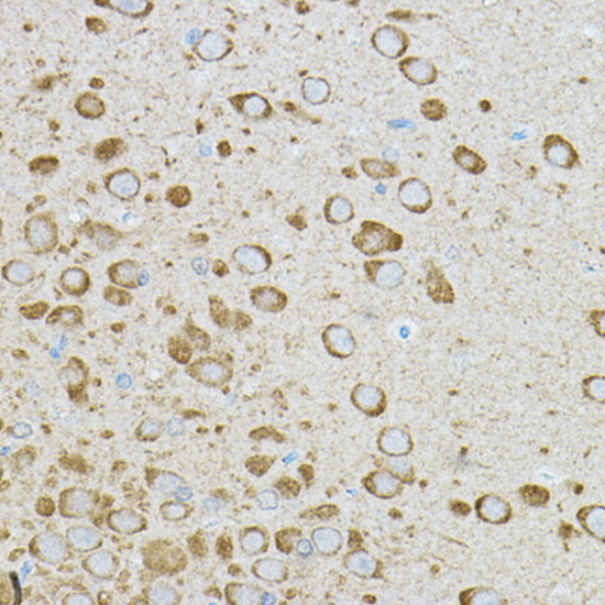
Immunohistochemistry analysis of paraffin-embedded Rat brain using IRAK4 Rabbit pAb (A6208) at dilution of 1:50 (40x lens). High pressure antigen retrieval performed with 0.01M Citrate buffer (pH 6.0) prior to IHC staining.
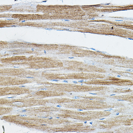
Immunohistochemistry analysis of paraffin-embedded Rat heart using IRAK4 Rabbit pAb (A6208) at dilution of 1:50 (40x lens). High pressure antigen retrieval performed with 0.01M Citrate buffer (pH 6.0) prior to IHC staining.
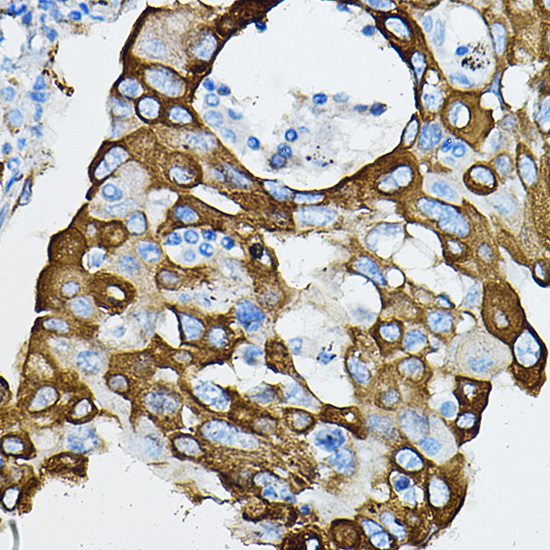
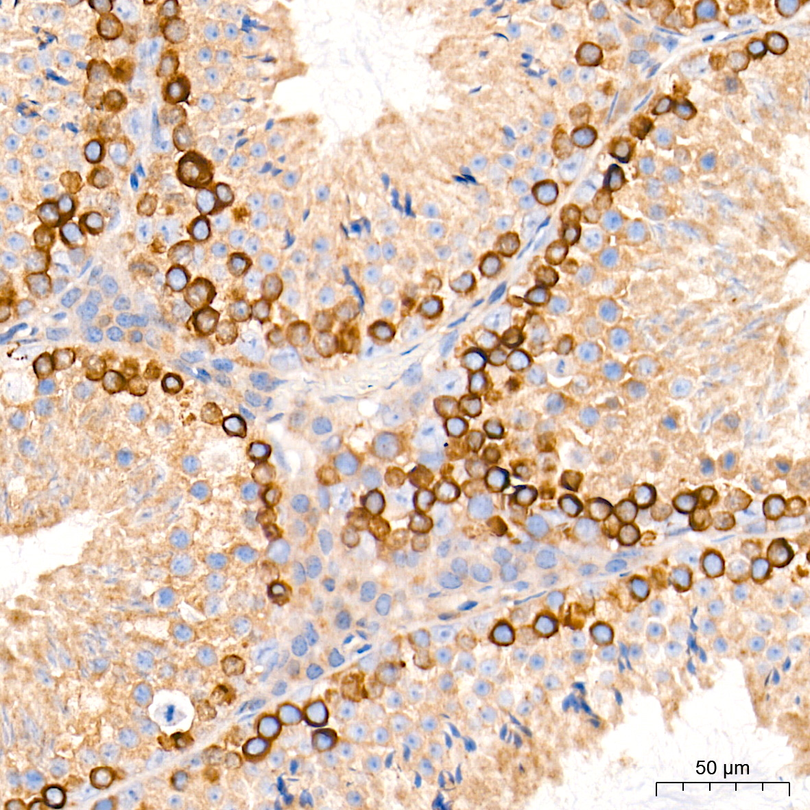
Immunohistochemistry analysis of paraffin-embedded Human lung cancer using IRAK4 Rabbit pAb (A6208) at dilution of 1:50 (40x lens). High pressure antigen retrieval performed with 0.01M Citrate buffer (pH 6.0) prior to IHC staining.
Immunohistochemistry analysis of paraffin-embedded Human hepatobiliary duct using IRAK4 Rabbit pAb (A6208) at dilution of 1:50 (40x lens). High pressure antigen retrieval performed with 0.01M Citrate buffer (pH 6.0) prior to IHC staining.
Immunofluorescence analysis of NIH/3T3 cells using IRAK4 Rabbit pAb (A6208) at dilution of 1:100 (40x lens). Secondary antibody: Cy3-conjugated Goat anti-Rabbit IgG (H+L) (AS007) at 1:500 dilution. Blue: DAPI for nuclear staining.
[KO Validated] TBK1/NAK Rabbit mAb
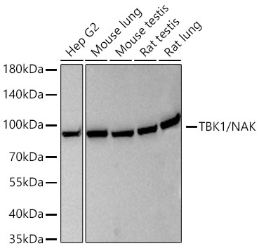
Western blot analysis of various lysates using [KO Validated] TBK1/NAK Rabbit mAb (A3458) at 1:4000 dilution incubated at room temperature for 1.5 hours.
Secondary antibody: HRP-conjugated Goat anti-Rabbit IgG (H+L) (AS014) at 1:10000 dilution.
Lysates/proteins: 25 μg per lane.
Blocking buffer: 3% nonfat dry milk in TBST.
Detection: ECL Basic Kit (RM00020).
Exposure time: 90 s.
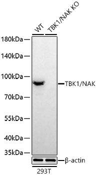
Western blot analysis of lysates from wild type (WT) and TBK1/NAK knockout (KO) 293T cells using [KO Validated] TBK1/NAK Rabbit mAb (A3458) at 1:7000 dilution incubated at room temperature for 1.5 hours.
Secondary antibody: HRP-conjugated Goat anti-Rabbit IgG (H+L) (AS014) at 1:10000 dilution.
Lysates/proteins: 25 μg per lane.
Blocking buffer: 3% nonfat dry milk in TBST.
Detection: ECL Basic Kit (RM00020).
Exposure time: 90 s.
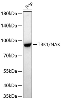
Western blot analysis of lysates from Raji cells using [KO Validated] TBK1/NAK Rabbit mAb (A3458) at 1:7000 dilution incubated at room temperature for 1.5 hours.
Secondary antibody: HRP-conjugated Goat anti-Rabbit IgG (H+L) (AS014) at 1:10000 dilution.
Lysates/proteins: 25 μg per lane.
Blocking buffer: 3% nonfat dry milk in TBST.
Detection: ECL Basic Kit (RM00020).
Exposure time: 90 s.
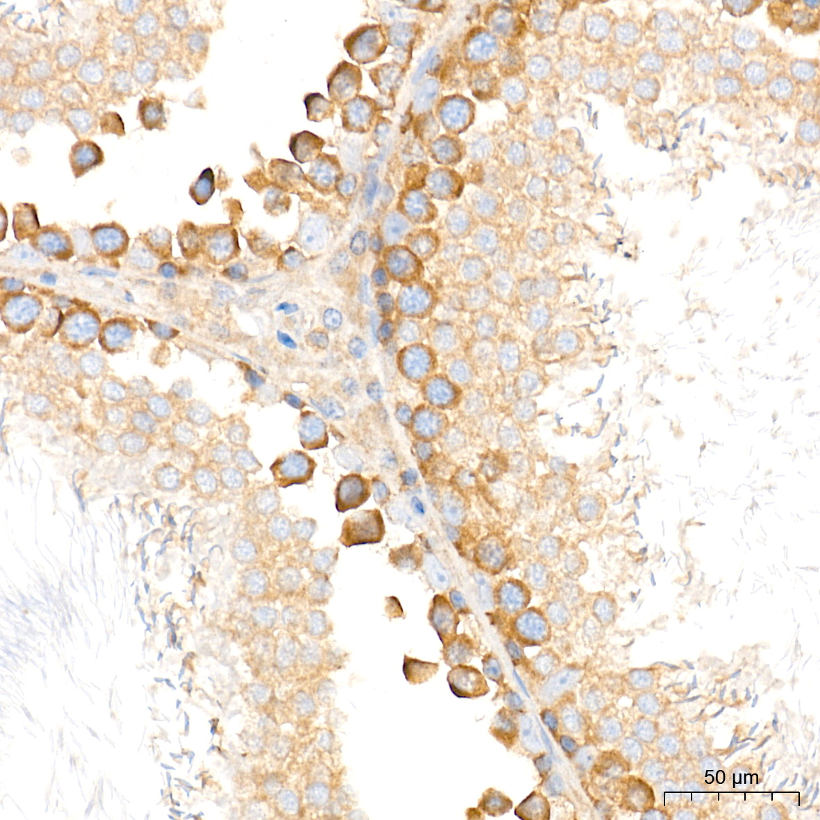
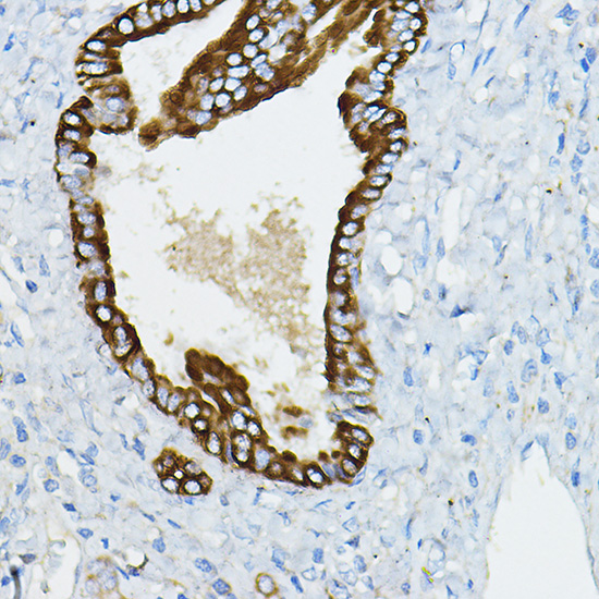
Immunohistochemistry analysis of paraffin-embedded Rat testis tissue using [KO Validated] TBK1/NAK Rabbit mAb (A3458) at a dilution of 1:750 (40x lens). High pressure antigen retrieval performed with 0.01M Tris-EDTA Buffer (pH 9.0) prior to IHC staining.
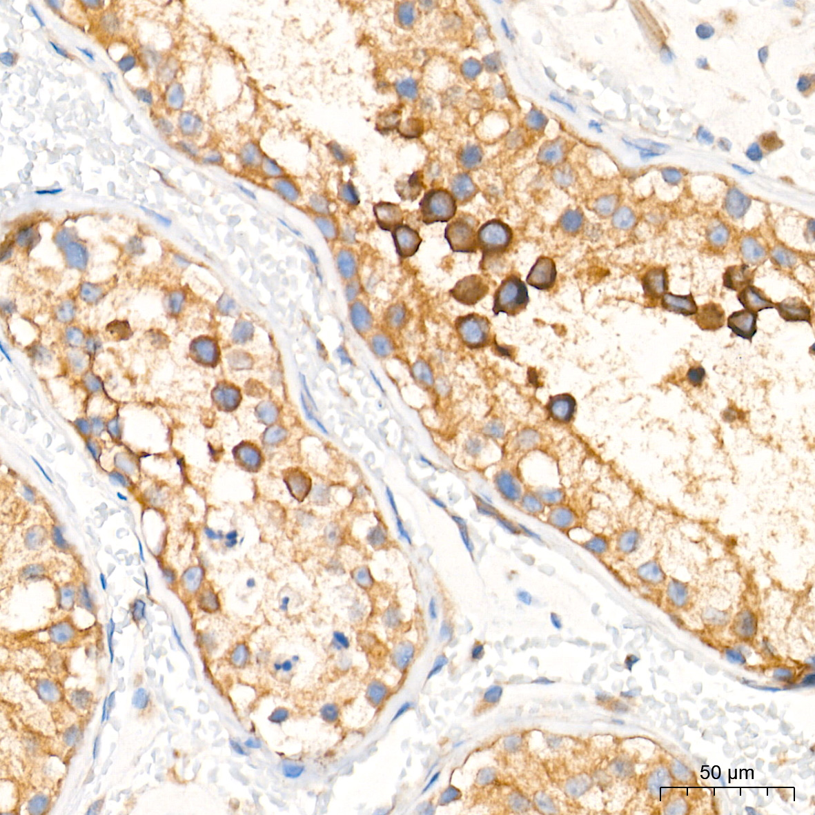
Immunohistochemistry analysis of paraffin-embedded Human testis tissue using [KO Validated] TBK1/NAK Rabbit mAb (A3458) at a dilution of 1:750 (40x lens). High pressure antigen retrieval performed with 0.01M Tris-EDTA Buffer (pH 9.0) prior to IHC staining.
Immunohistochemistry analysis of paraffin-embedded Mouse testis tissue using [KO Validated] TBK1/NAK Rabbit mAb (A3458) at a dilution of 1:750 (40x lens). High pressure antigen retrieval performed with 0.01M Tris-EDTA Buffer (pH 9.0) prior to IHC staining.
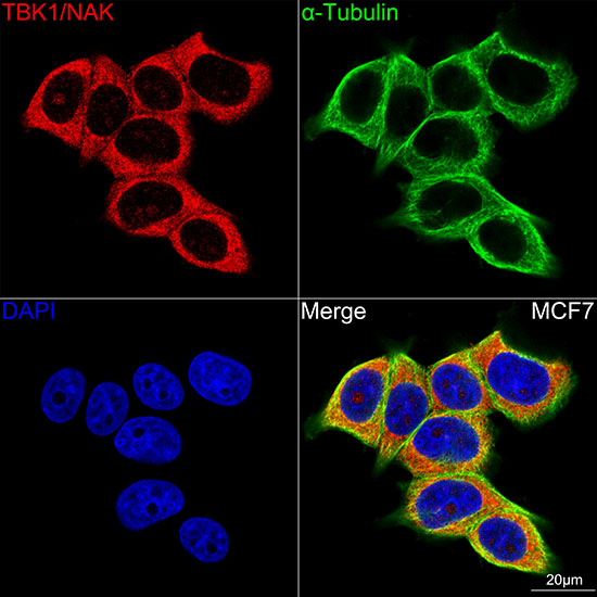
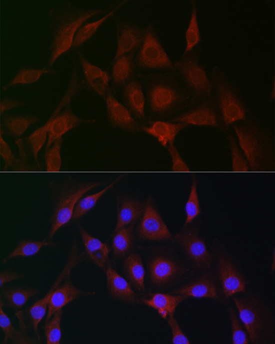
Confocal imaging of MCF7 cells using [KO Validated] TBK1/NAK Rabbit mAb (A3458,at dilution of 1:100) (Red). The cells were counterstained with α-Tubulin Mouse mAb (AC012,dilution 1:400) (Green). DAPI was used for nuclear staining (blue). Objective: 100x.
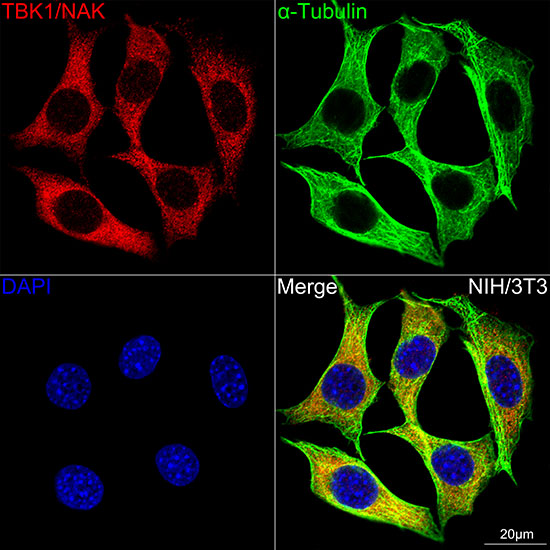
Confocal imaging of NIH/3T3 cells using [KO Validated] TBK1/NAK Rabbit mAb (A3458,at dilution of 1:100) (Red). The cells were counterstained with α-Tubulin Mouse mAb (AC012,dilution 1:400) (Green). DAPI was used for nuclear staining (blue). Objective: 100x.
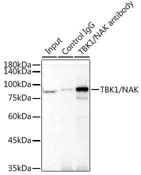
Immunoprecipitation analysis of 300 μg extracts from 293T cells using 3 μg [KO Validated] TBK1/NAK Rabbit mAb (A3458). Western blot was performed from the immunoprecipitate using [KO Validated] TBK1/NAK Rabbit mAb (A3458) at a dilution of 1:1000.
Phospho-TBK1/NAK-S172 Rabbit mAb
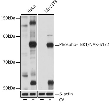
Western blot analysis of various lysates using Phospho-TBK1/NAK-S172 Rabbit mAb (AP1026) at 1:1000 dilution. Both HeLa cells and NIH/3T3 cells were treated with Calyculin A (100 nM) at 37℃ for 30 minutes after serum-starvation overnight.
Secondary antibody: HRP-conjugated Goat anti-Rabbit IgG (H+L) (AS014) at 1:10000 dilution.
Lysates/proteins: 25μg per lane.
Blocking buffer: 3% BSA.
Detection: ECL Basic Kit (RM00020).
Exposure time: 1min.
Phospho-JNK1/2/3-T183/T183/T221 Rabbit mAb
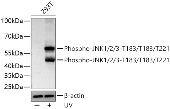
Western blot analysis of lysates from 293T cells using Phospho-JNK1/2/3-T183/T183/T221 Rabbit mAb (AP0631) at 1:1000 dilution incubated overnight at 4℃. 293T cells were treated with UV (90mJ/cm2) at 37°C for 30min
Secondary antibody: HRP-conjugated Goat anti-Rabbit IgG (H+L) (AS014) at 1:10000 dilution.
Lysates/proteins: μg per lane.
Blocking buffer: 3% nonfat dry milk in TBST.
Detection: ECL Basic Kit (RM00020).
Exposure time: 90 s.
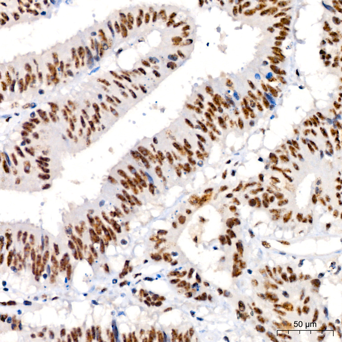
Immunohistochemistry analysis of paraffin-embedded Human colon carcinoma using Phospho-JNK1/2/3-T183/T183/T221 Rabbit mAb (AP0631) at dilution of 1:200 (40x lens). High pressure antigen retrieval performed with 0.01M Citrate buffer (pH 6.0) prior to IHC staining.
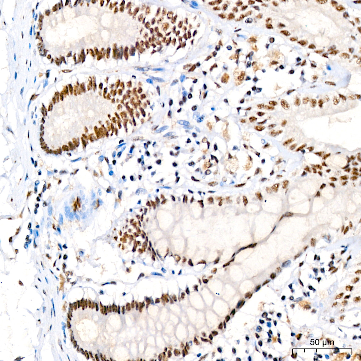
Immunohistochemistry analysis of paraffin-embedded Human colon using Phospho-JNK1/2/3-T183/T183/T221 Rabbit mAb (AP0631) at dilution of 1:200 (40x lens). High pressure antigen retrieval performed with 0.01M Citrate buffer (pH 6.0) prior to IHC staining.
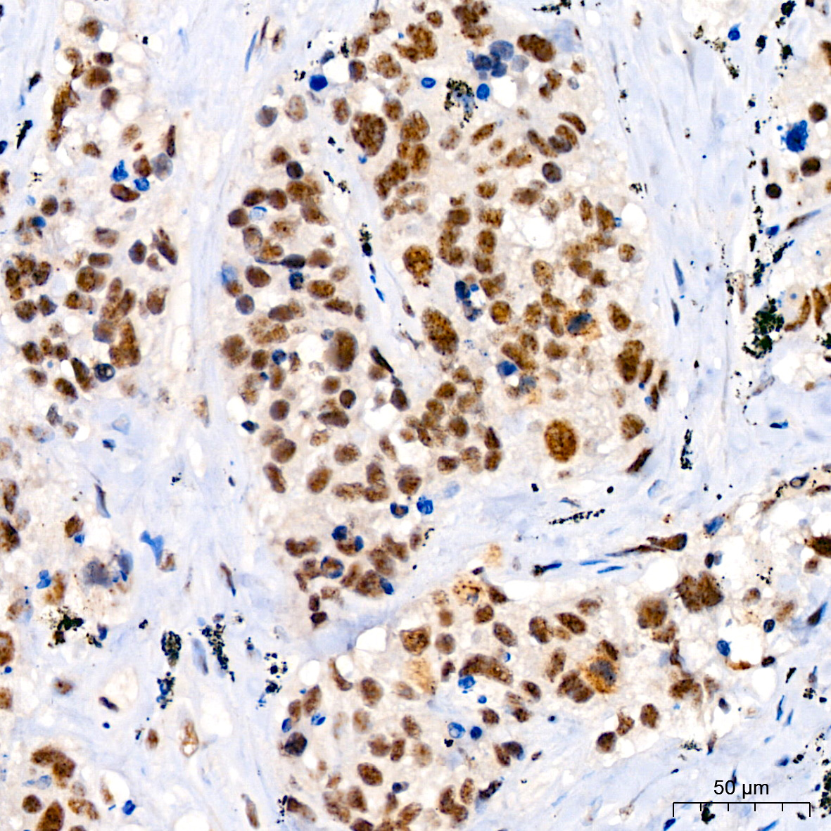
Immunohistochemistry analysis of paraffin-embedded Human lung squamous carcinoma tissue using Phospho-JNK1/2/3-T183/T183/T221 Rabbit mAb (AP0631) at dilution of 1:200 (40x lens). High pressure antigen retrieval performed with 0.01M Citrate buffer (pH 6.0) prior to IHC staining.
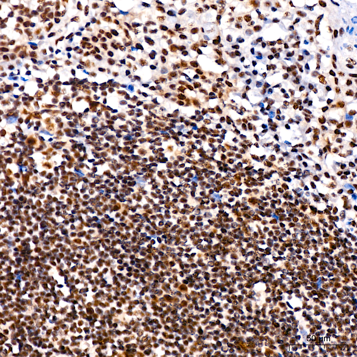
Immunohistochemistry analysis of paraffin-embedded Human tonsil using Phospho-JNK1/2/3-T183/T183/T221 Rabbit mAb (AP0631) at dilution of 1:200 (40x lens). High pressure antigen retrieval performed with 0.01M Citrate buffer (pH 6.0) prior to IHC staining.
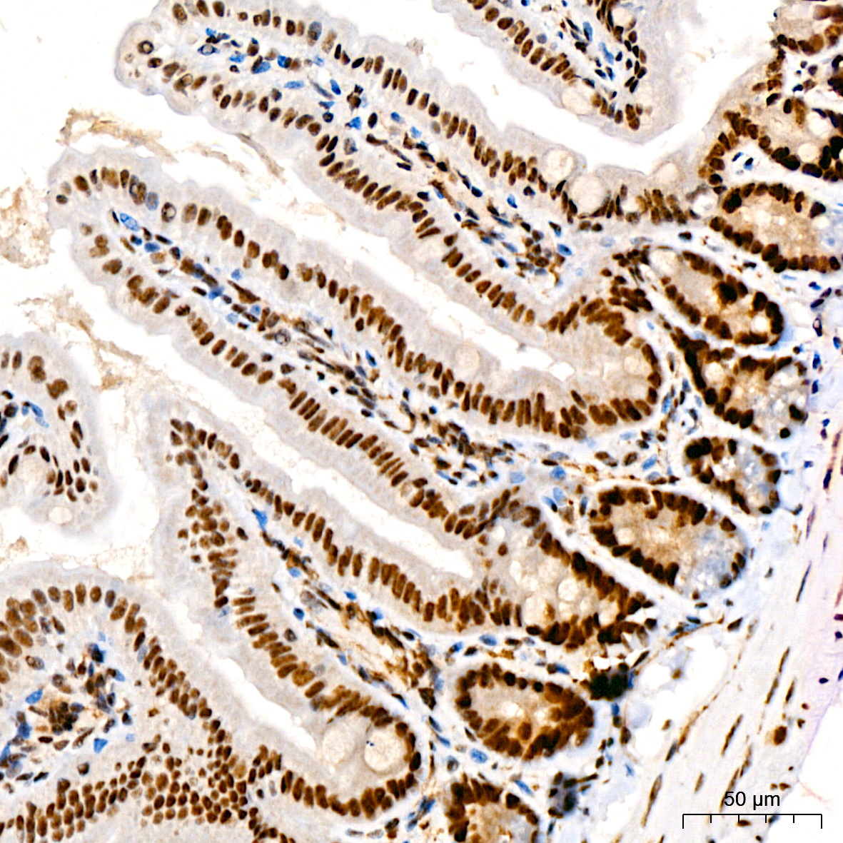
Immunohistochemistry analysis of paraffin-embedded Mouse colon using Phospho-JNK1/2/3-T183/T183/T221 Rabbit mAb (AP0631) at dilution of 1:200 (40x lens). High pressure antigen retrieval performed with 0.01M Citrate buffer (pH 6.0) prior to IHC staining.
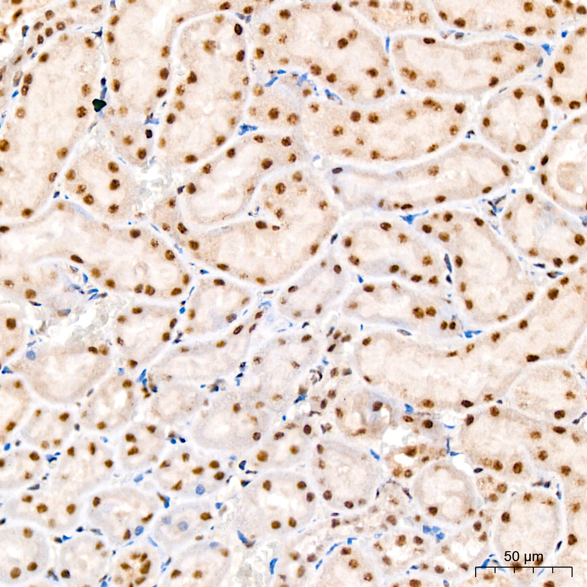
Immunohistochemistry analysis of paraffin-embedded Rat kidney using Phospho-JNK1/2/3-T183/T183/T221 Rabbit mAb (AP0631) at dilution of 1:200 (40x lens). High pressure antigen retrieval performed with 0.01M Citrate buffer (pH 6.0) prior to IHC staining.
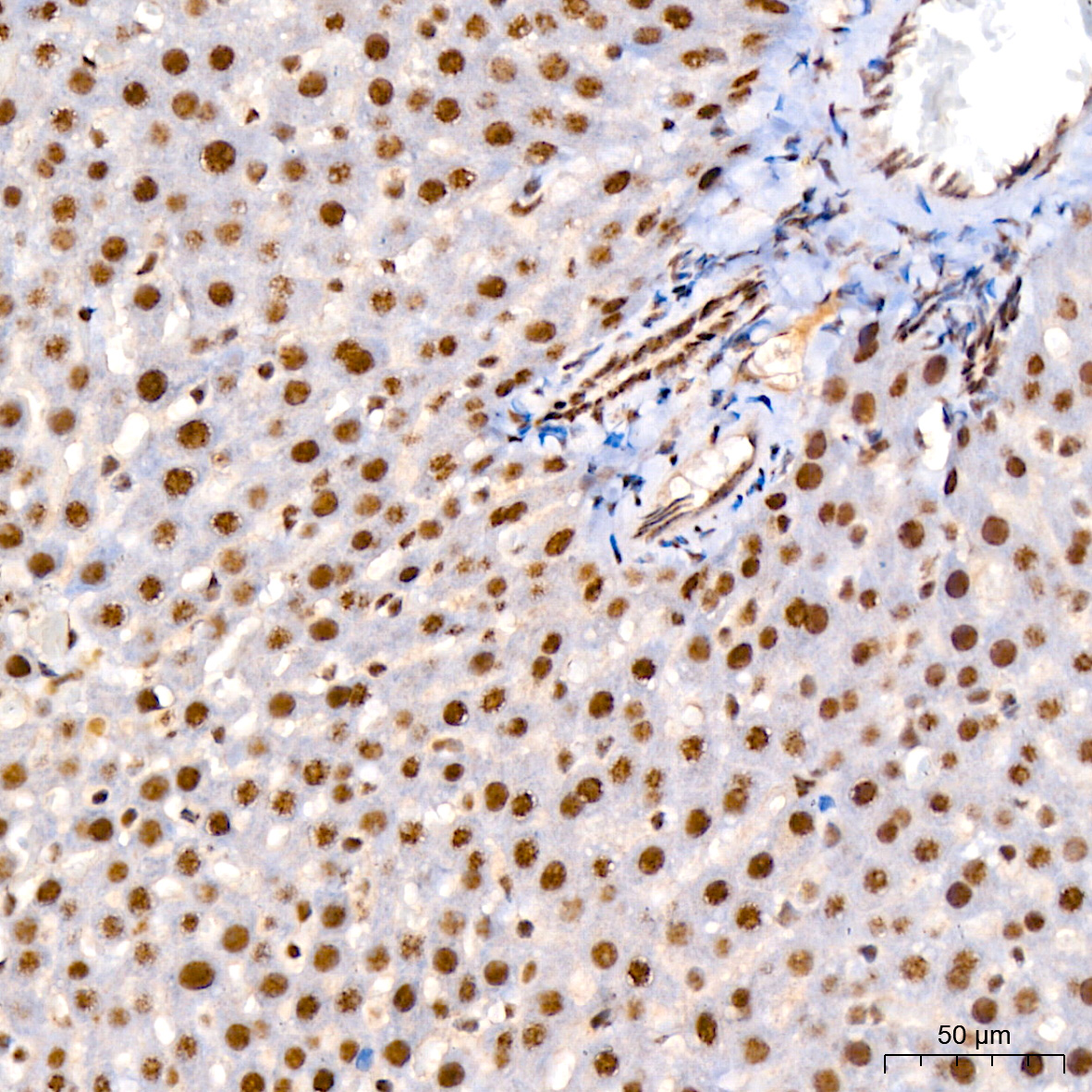
Immunohistochemistry analysis of paraffin-embedded Rat liver using Phospho-JNK1/2/3-T183/T183/T221 Rabbit mAb (AP0631) at dilution of 1:200 (40x lens). High pressure antigen retrieval performed with 0.01M Citrate buffer (pH 6.0) prior to IHC staining.
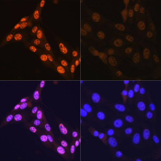
Immunofluorescence analysis of NIH-3T3 cells using Phospho-JNK1/2/3-T183/T183/T221 Rabbit mAb (AP0631).NIH-3T3 cells were treated with Anisomycin (25 μg/mL) at 37℃ for 30 minutes after serum-starvation overnight. Secondary antibody: Cy3-conjugated Goat anti-Rabbit IgG (H+L) (AS007) at 1:500 dilution. Blue: DAPI for nuclear staining.