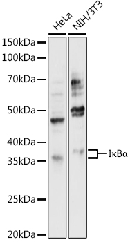
Western blot analysis of various lysates using IκBα Rabbit pAb (A11397) at 1:1000 dilution.
Secondary antibody: HRP-conjugated Goat anti-Rabbit IgG (H+L) (AS014) at 1:10000 dilution.
Lysates/proteins: 25μg per lane.
Blocking buffer: 3% nonfat dry milk in TBST.
Detection: ECL Enhanced Kit (RM00021).
Exposure time: 180s.
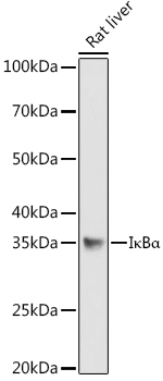
Western blot analysis of lysates from Rat liver, using IκBα Rabbit pAb (A11397) at 1:1000 dilution.
Secondary antibody: HRP-conjugated Goat anti-Rabbit IgG (H+L) (AS014) at 1:10000 dilution.
Lysates/proteins: 25μg per lane.
Blocking buffer: 3% nonfat dry milk in TBST.
Detection: ECL Basic Kit (RM00020).
Exposure time: 180s.
[KO Validated] NF-kB p65/RelA Rabbit mAb
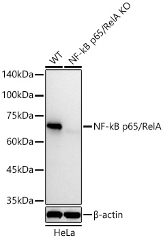
Western blot analysis of lysates from wild type (WT) and NF-kB p65/RelA knockout (KO) HeLa cells using [KO Validated] NF-kB p65/RelA Rabbit mAb (A19653) at 1:10000 dilution incubated overnight at 4℃.
Secondary antibody: HRP-conjugated Goat anti-Rabbit IgG (H+L) (AS014) at 1:10000 dilution.
Lysates/proteins: 25 μg per lane.
Blocking buffer: 3% nonfat dry milk in TBST.
Detection: ECL Basic Kit (RM00020).
Exposure time: 30s.
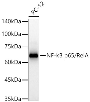
Western blot analysis of lysates from PC-12 cells using [KO Validated] NF-kB p65/RelA Rabbit mAb (A19653) at 1:10000 dilution incubated overnight at 4℃.
Secondary antibody: HRP-conjugated Goat anti-Rabbit IgG (H+L) (AS014) at 1:10000 dilution.
Lysates/proteins: 25 μg per lane.
Blocking buffer: 3% nonfat dry milk in TBST.
Detection: ECL Basic Kit (RM00020).
Exposure time: 30s.
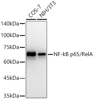
Western blot analysis of various lysates using [KO Validated] NF-kB p65/RelA Rabbit mAb (A19653) at 1:10000 dilution incubated overnight at 4℃.
Secondary antibody: HRP-conjugated Goat anti-Rabbit IgG (H+L) (AS014) at 1:10000 dilution.
Lysates/proteins: 25 μg per lane.
Blocking buffer: 3% nonfat dry milk in TBST.
Detection: ECL Basic Kit (RM00020).
Exposure time: 60s.
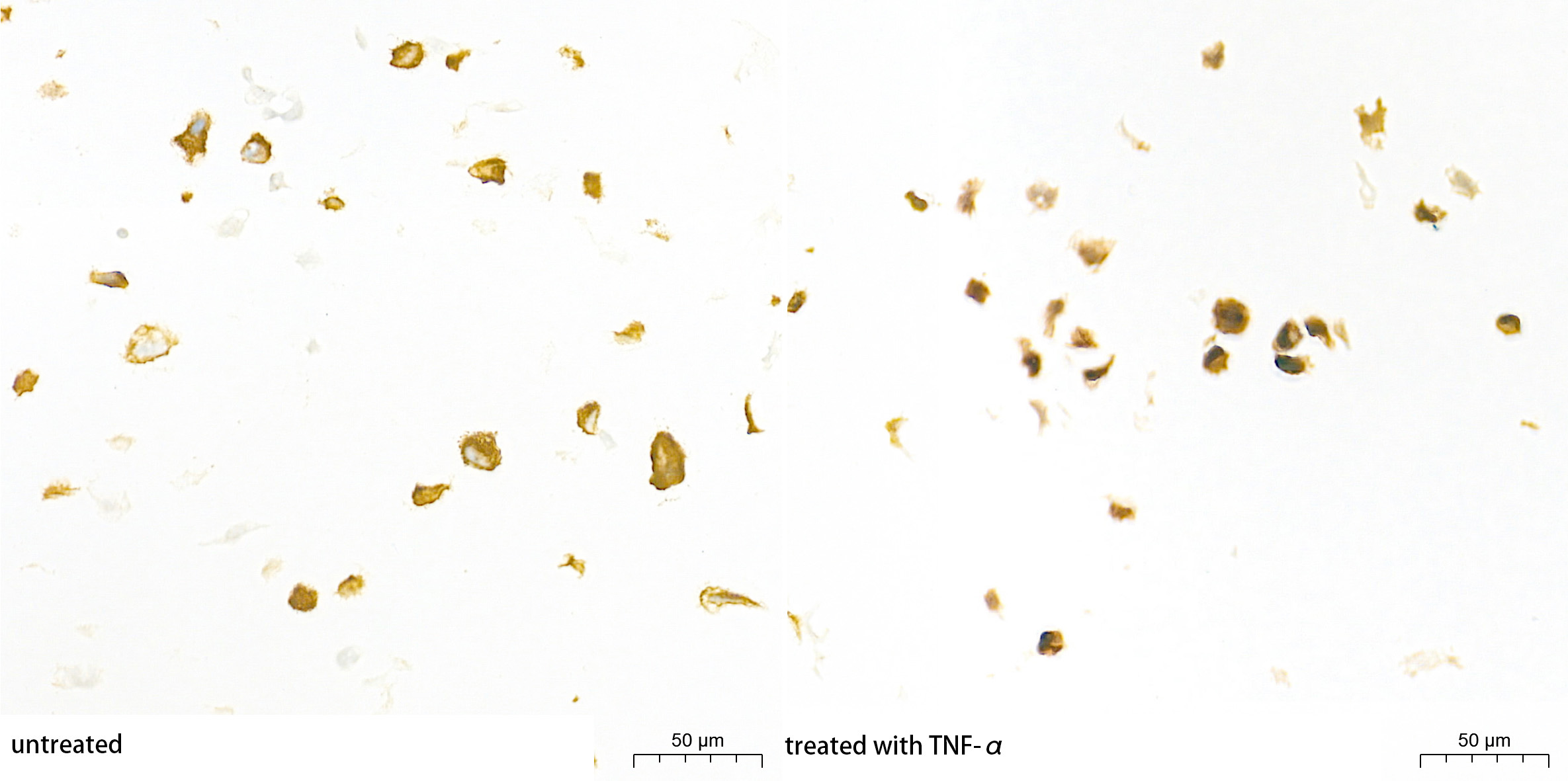
Immunohistochemistry analysis of paraffin-embedded HT-1080 cell lines(untreated and treated with TNF-α) using [KO Validated] NF-kB p65/RelA Rabbit mAb (A19653) at a dilution of 1:3000 (40x lens). High pressure antigen retrieval performed with 0.01M Tris-EDTA Buffer (pH 9.0) prior to IHC staining.
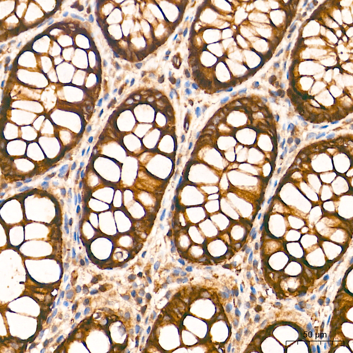
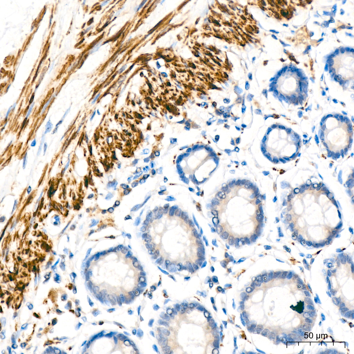
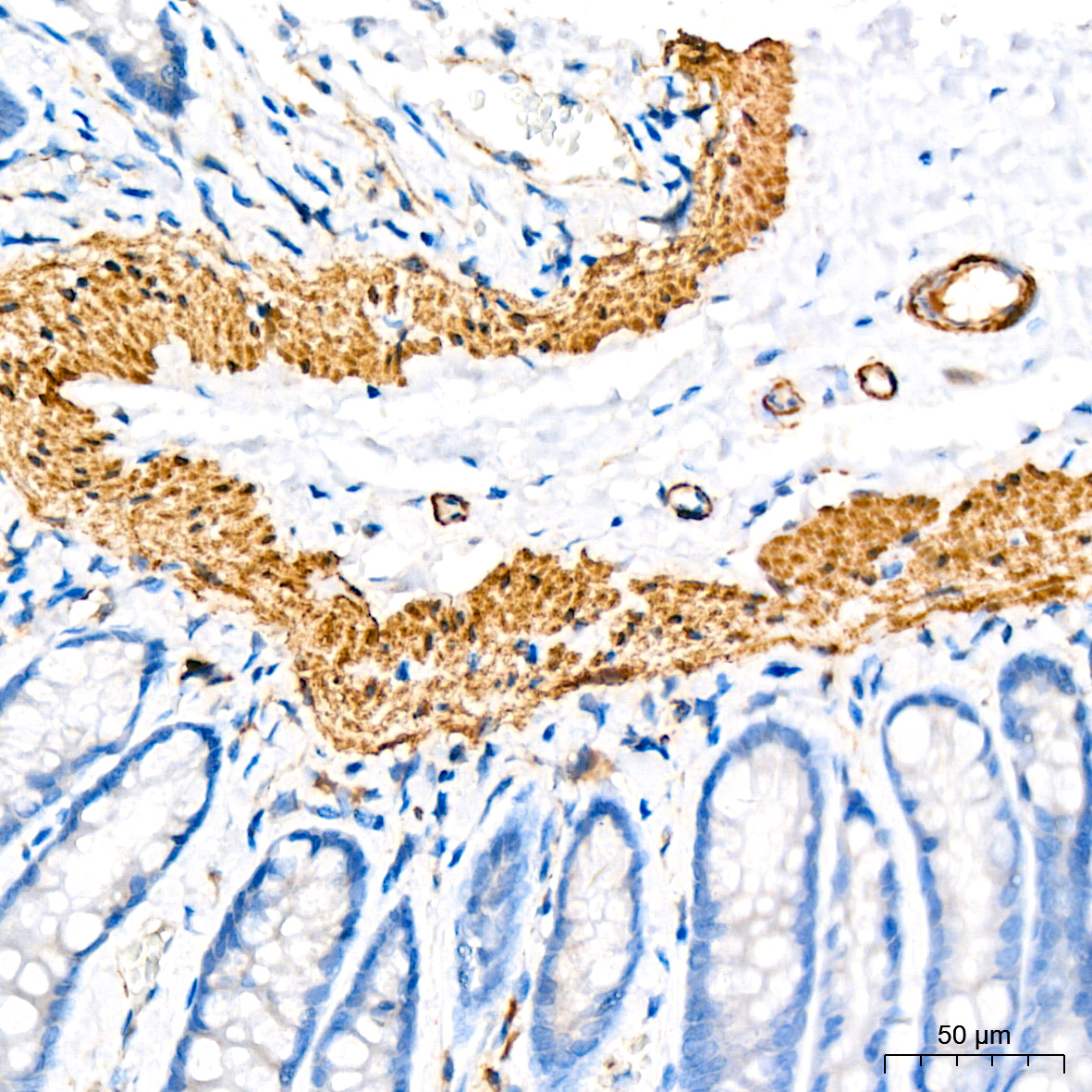
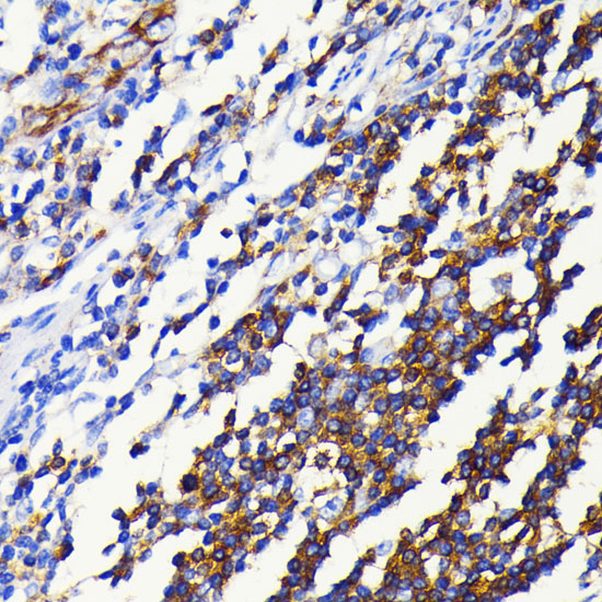
Immunohistochemistry analysis of paraffin-embedded Human colon tissue using [KO Validated] NF-kB p65/RelA Rabbit mAb (A19653) at a dilution of 1:3000 (40x lens). High pressure antigen retrieval performed with 0.01M Tris-EDTA Buffer (pH 9.0) prior to IHC staining.
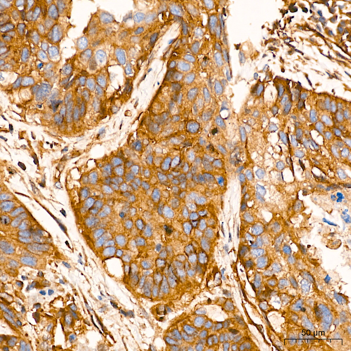
Immunohistochemistry analysis of paraffin-embedded Human lung cancer tissue using [KO Validated] NF-kB p65/RelA Rabbit mAb (A19653) at a dilution of 1:3000 (40x lens). High pressure antigen retrieval performed with 0.01M Tris-EDTA Buffer (pH 9.0) prior to IHC staining.
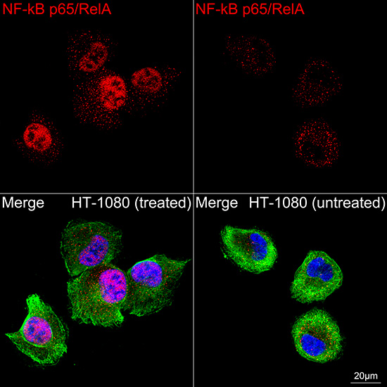
Confocal imaging of HT-1080 cells (treated with TNF-α) and HT-1080 cells (untreated) cells using [KO Validated] NF-kB p65/RelA Rabbit mAb (A19653, dilution 1:2100) followed by a further incubation with Cy3 Goat Anti-Rabbit IgG (H+L) (AS007, dilution 1:500) (Red). The cells were counterstained with α-Tubulin Mouse mAb (AC012, dilution 1:400) followed by incubation with ABflo® 488-conjugated Goat Anti-Mouse IgG (H+L) Ab (AS076, dilution 1:500) (Green). DAPI was used for nuclear staining (Blue). Objective: 100x.
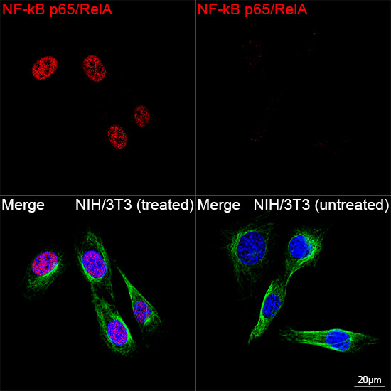
Confocal imaging of NIH/3T3 cells (treated with TNF-α) and NIH/3T3 cells (untreated) cells using [KO Validated] NF-kB p65/RelA Rabbit mAb (A19653, dilution 1:2100) followed by a further incubation with Cy3 Goat Anti-Rabbit IgG (H+L) (AS007, dilution 1:500) (Red). The cells were counterstained with α-Tubulin Mouse mAb (AC012, dilution 1:400) followed by incubation with ABflo® 488-conjugated Goat Anti-Mouse IgG (H+L) Ab (AS076, dilution 1:500) (Green). DAPI was used for nuclear staining (Blue). Objective: 100x.
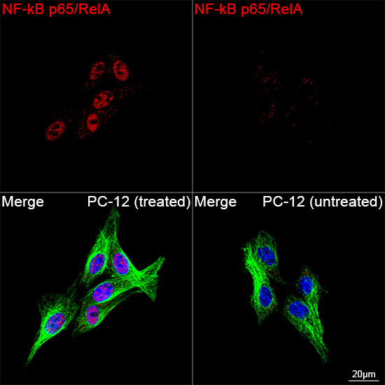
Confocal imaging of PC-12 cells (treated with TNF-α) and PC-12 cells (untreated) cells using [KO Validated] NF-kB p65/RelA Rabbit mAb (A19653, dilution 1:2100) followed by a further incubation with Cy3 Goat Anti-Rabbit IgG (H+L) (AS007, dilution 1:500) (Red). The cells were counterstained with α-Tubulin Mouse mAb (AC012, dilution 1:400) followed by incubation with ABflo® 488-conjugated Goat Anti-Mouse IgG (H+L) Ab (AS076, dilution 1:500) (Green). DAPI was used for nuclear staining (Blue). Objective: 100x.
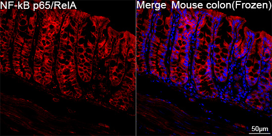
Confocal imaging of frozen sections Mouse colon tissue using [KO Validated] NF-kB p65/RelA Rabbit mAb (A19653, dilution 1:200) followed by a further incubation with Cy3-conjugated Goat anti-Rabbit IgG (H+L) (AS007, dilution 1:500) (Red). DAPI was used for nuclear staining (Blue). Microwave antigen retrieval performed with 0.01M Citrate Buffer (pH 6.0) prior to IF staining. Objective: 40x.
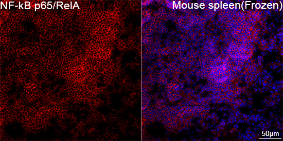
Confocal imaging of frozen sections Mouse spleen tissue using [KO Validated] NF-kB p65/RelA Rabbit mAb (A19653, dilution 1:200) followed by a further incubation with Cy3-conjugated Goat anti-Rabbit IgG (H+L) (AS007, dilution 1:500) (Red). DAPI was used for nuclear staining (Blue). Microwave antigen retrieval performed with 0.01M Citrate Buffer (pH 6.0) prior to IF staining. Objective: 40x.
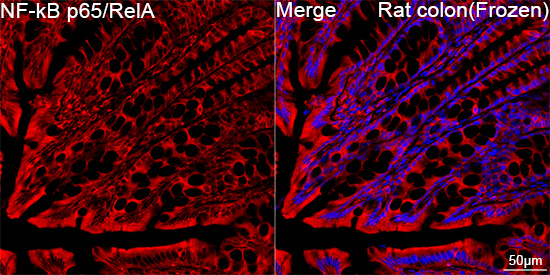
Confocal imaging of frozen sections Rat colon tissue using [KO Validated] NF-kB p65/RelA Rabbit mAb (A19653, dilution 1:200) followed by a further incubation with Cy3-conjugated Goat anti-Rabbit IgG (H+L) (AS007, dilution 1:500) (Red). DAPI was used for nuclear staining (Blue). Microwave antigen retrieval performed with 0.01M Citrate Buffer (pH 6.0) prior to IF staining. Objective: 40x.
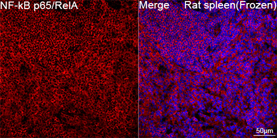
Confocal imaging of frozen sections Rat spleen tissue using [KO Validated] NF-kB p65/RelA Rabbit mAb (A19653, dilution 1:200) followed by a further incubation with Cy3-conjugated Goat anti-Rabbit IgG (H+L) (AS007, dilution 1:500) (Red). DAPI was used for nuclear staining (Blue). Microwave antigen retrieval performed with 0.01M Citrate Buffer (pH 6.0) prior to IF staining. Objective: 40x.
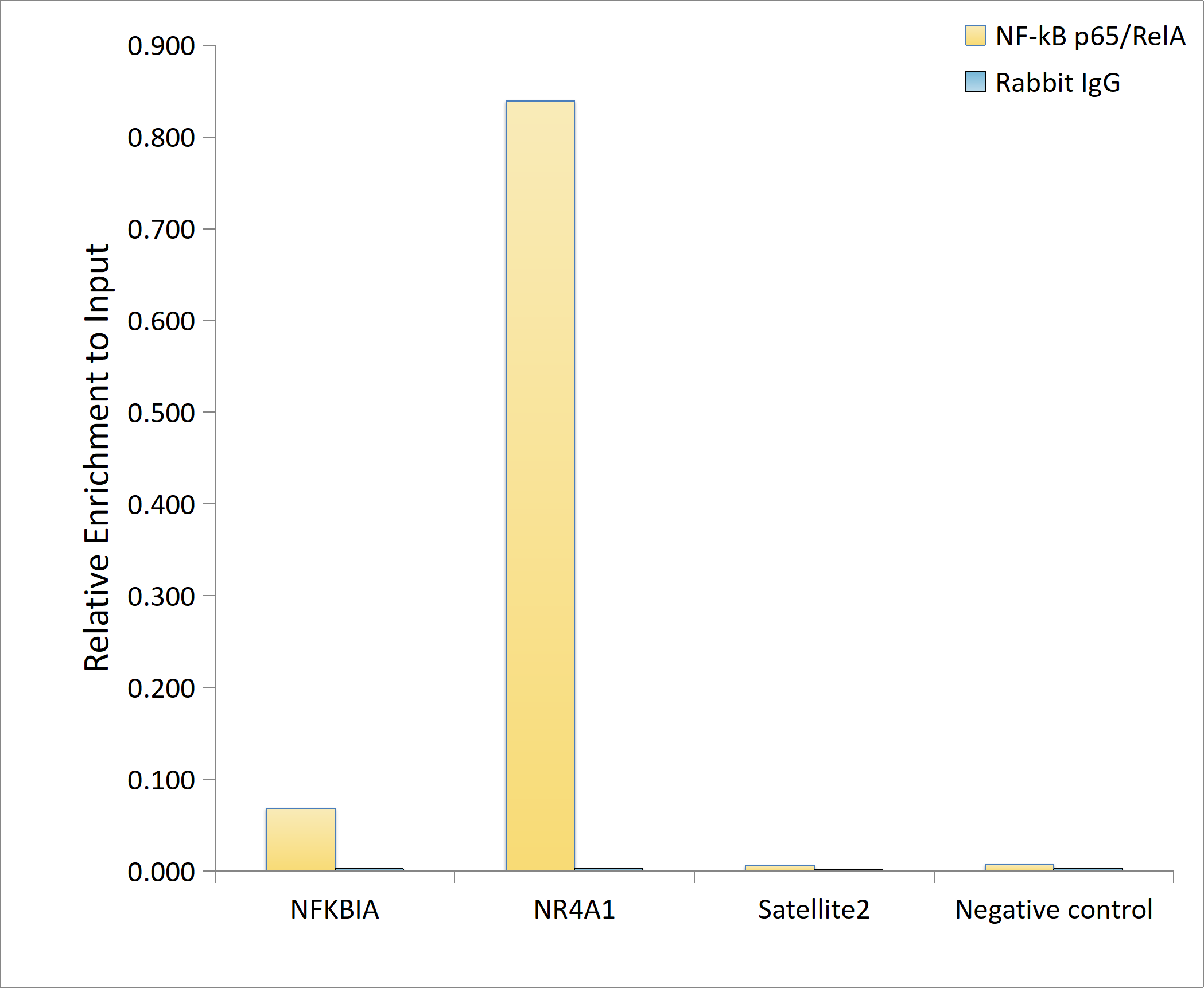
Chromatin immunoprecipitation was performed with 10 μg of cross-linked chromatin from HT-1080 cells treated by TNF-α (20 ng/ml) at 37℃ for 30 minutes, using 5 μg of [KO Validated] NF-kB p65/RelA Rabbit mAb (A19653) and Rabbit IgG isotype control (AC042). The enrichment of immunoprecipitated DNA at different genomic loci was examined by quantitative PCR. The histogram compares the ratio of the immunoprecipitated DNA to the input at given loci.
Phospho-NF-kB p65/RelA-S536 Rabbit mAb
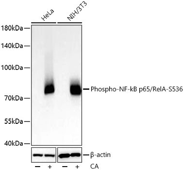
Western blot analysis of various lysates using Phospho-NF-kB p65/RelA-S536 Rabbit mAb (AP1294) at 1:10000 dilution incubated overnight at 4℃. HeLa cells were treated with CA (100nM) at 37°C for 30min, NIH/3T3 cells were treated with CA (100nM) at 37°C for 30min
Secondary antibody: HRP-conjugated Goat anti-Rabbit IgG (H+L) (AS014) at 1:10000 dilution.
Lysates/proteins: 30 μg per lane.
Blocking buffer: 3% nonfat dry milk in TBST.
Detection: ECL Basic Kit (RM00020).
Exposure time: 20 s.
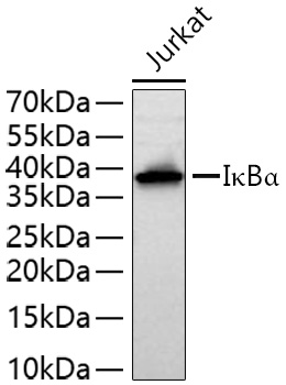
Western blot analysis of lysates from Jurkat cells using IκBα Rabbit mAb (A19714) at 1:4000 dilution incubated overnight at 4℃.
Secondary antibody: HRP-conjugated Goat anti-Rabbit IgG (H+L) (AS014) at 1:10000 dilution.
Lysates/proteins: 25 μg per lane.
Blocking buffer: 3% nonfat dry milk in TBST.
Detection: ECL Basic Kit (RM00020).
Exposure time: 20 s.
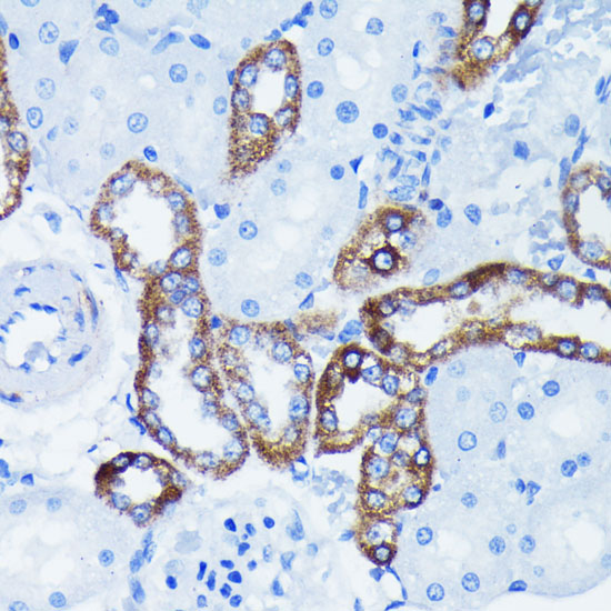
Immunohistochemistry analysis of paraffin-embedded Rat kidney using IκBα Rabbit mAb (A19714) at dilution of 1:100 (40x lens). Microwave antigen retrieval performed with 0.01M PBS Buffer (pH 7.2) prior to IHC staining.
Immunohistochemistry analysis of paraffin-embedded Human colon using IκBα Rabbit mAb (A19714) at dilution of 1:100 (40x lens). Microwave antigen retrieval performed with 0.01M PBS Buffer (pH 7.2) prior to IHC staining.
Immunohistochemistry analysis of paraffin-embedded Mouse kidney using IκBα Rabbit mAb (A19714) at dilution of 1:100 (40x lens). Microwave antigen retrieval performed with 0.01M PBS Buffer (pH 7.2) prior to IHC staining.
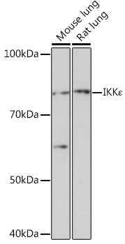
Western blot analysis of various lysates using IKKε Rabbit mAb (A3463) at 1:1000 dilution.
Secondary antibody: HRP-conjugated Goat anti-Rabbit IgG (H+L) (AS014) at 1:10000 dilution.
Lysates/proteins: 25μg per lane.
Blocking buffer: 3% nonfat dry milk in TBST.
Detection: ECL Basic Kit (RM00020).
Exposure time: 30s.
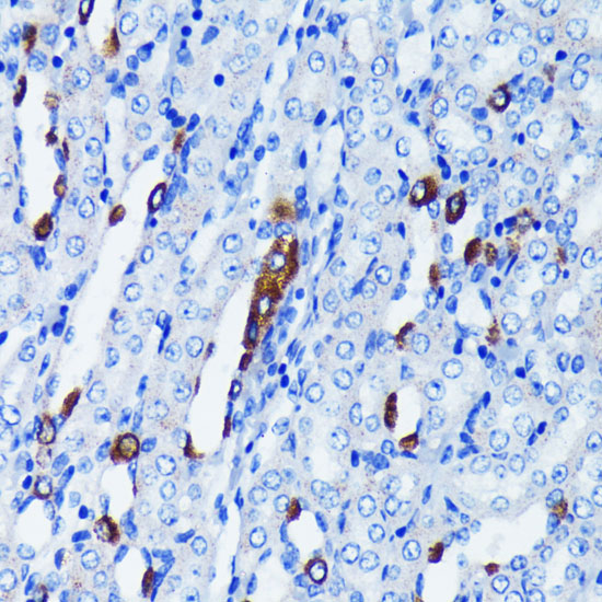
Immunohistochemistry analysis of paraffin-embedded Mouse heart using IKKε Rabbit mAb (A3463) at dilution of 1:100 (40x lens). Microwave antigen retrieval performed with 0.01M PBS Buffer (pH 7.2) prior to IHC staining.
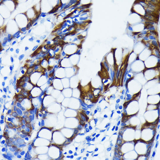
Immunohistochemistry analysis of paraffin-embedded Human colon tissue using IKKε Rabbit mAb (A3463) at a dilution of 1:200 (40x lens). High pressure antigen retrieval performed with 0.01M Citrate buffer (pH 6.0) prior to IHC staining.
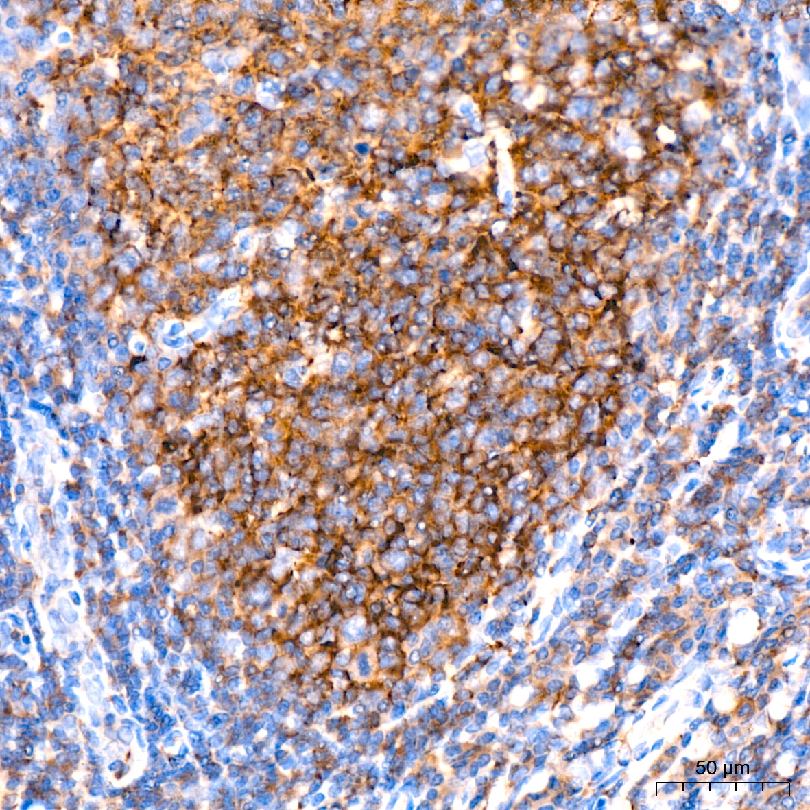
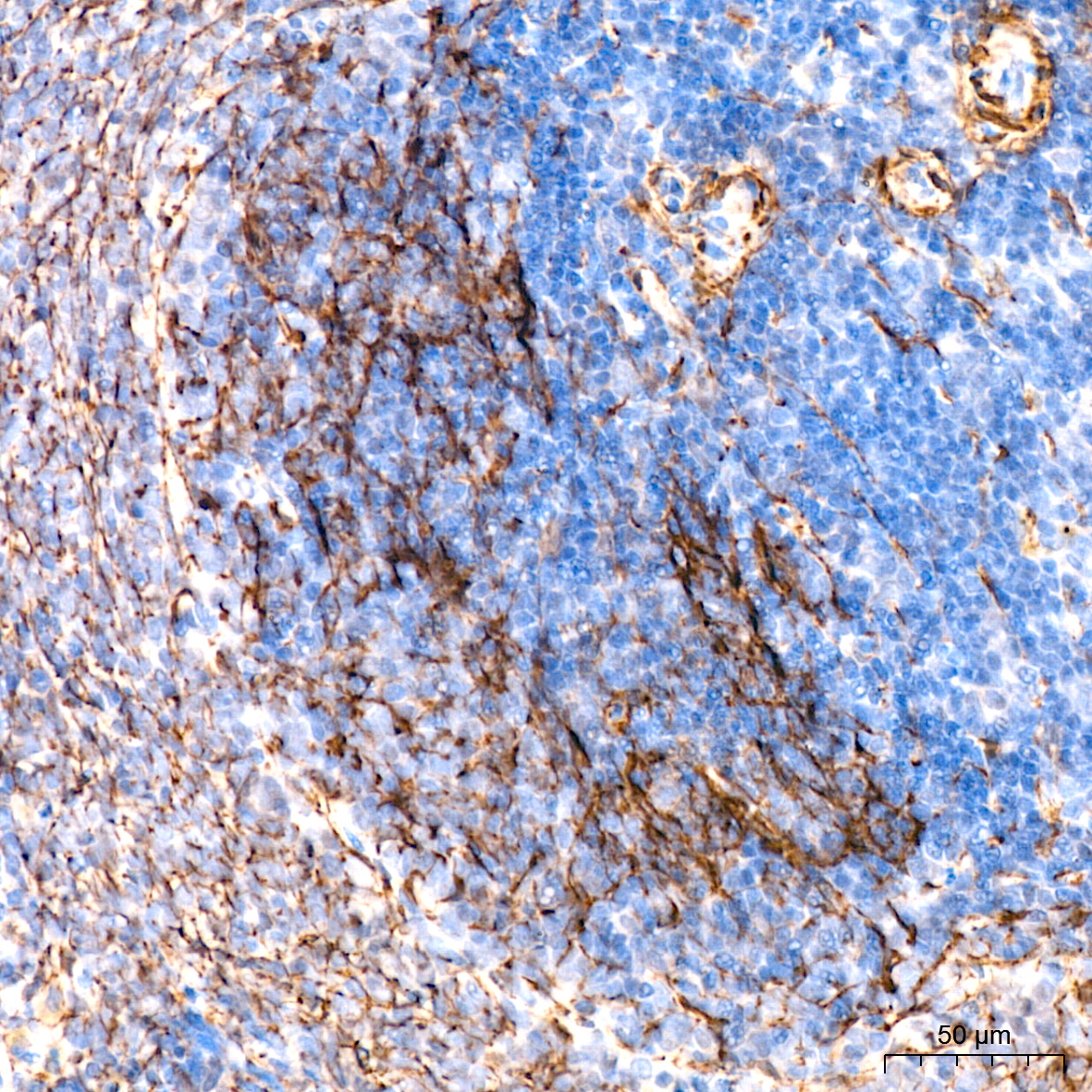
Immunohistochemistry analysis of paraffin-embedded Human tonsil tissue using IKKε Rabbit mAb (A3463) at a dilution of 1:200 (40x lens). High pressure antigen retrieval performed with 0.01M Citrate buffer (pH 6.0) prior to IHC staining.
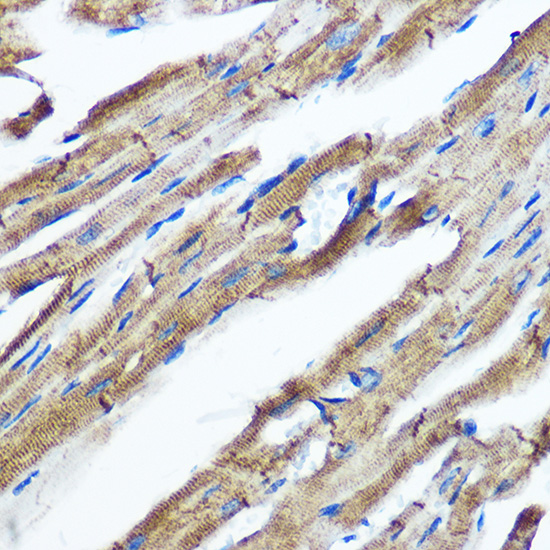
Immunohistochemistry analysis of paraffin-embedded Rat colon tissue using IKKε Rabbit mAb (A3463) at a dilution of 1:200 (40x lens). High pressure antigen retrieval performed with 0.01M Citrate buffer (pH 6.0) prior to IHC staining.
Immunohistochemistry analysis of paraffin-embedded Rat spleen tissue using IKKε Rabbit mAb (A3463) at a dilution of 1:200 (40x lens). High pressure antigen retrieval performed with 0.01M Citrate buffer (pH 6.0) prior to IHC staining.
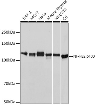
Western blot analysis of various lysates using NF-κB2 Rabbit mAb (A19605) at 1:1000 dilution.
Secondary antibody: HRP-conjugated Goat anti-Rabbit IgG (H+L) (AS014) at 1:10000 dilution.
Lysates/proteins: 25μg per lane.
Blocking buffer: 3% nonfat dry milk in TBST.
Detection: ECL Basic Kit (RM00020).
Exposure time: 3s.
Immunohistochemistry analysis of paraffin-embedded Human appendix using NF-κB2 Rabbit mAb (A19605) at dilution of 1:100 (40x lens). Microwave antigen retrieval performed with 0.01M PBS Buffer (pH 7.2) prior to IHC staining.