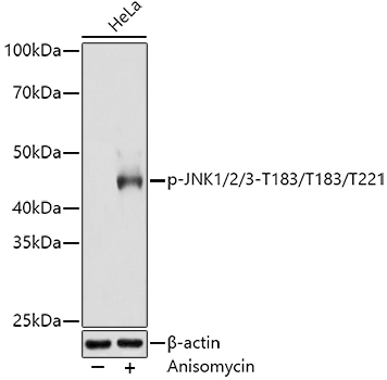
Western blot analysis of lysates from HeLa cells, using Phospho-JNK1/2/3-T183/T183/T221 Rabbit mAb (AP0631) at 1:3000 dilution. HeLa cells were treated by Anisomycin (25 μg/mL) at 37℃ for 30 minutes after serum-starvation overnight.Secondary antibody: HRP-conjugated Goat anti-Rabbit IgG (H+L) (AS014) at 1:10000 dilution.Lysates/proteins: 25μg per lane.Blocking buffer: 3% nonfat dry milk in TBST.Detection: ECL Basic Kit (RM00020).Exposure time: 180s.
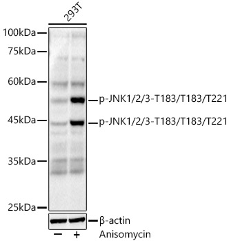
Western blot analysis of lysates from 293T cells, using Phospho-JNK1/2/3-T183/T183/T221 Rabbit mAb (A23099) at 1:1000 dilution. 293T cells were treated by Anisomycin (25 μg/mL) at 37℃ for 30 minutes after serum-starvation overnight.Secondary antibody: HRP-conjugated Goat anti-Rabbit IgG (H+L) (AS014) at 1:10000 dilution.Lysates/proteins: 25μg per lane.Blocking buffer: 3% nonfat dry milk in TBST.Detection: ECL Basic Kit (RM00020).Exposure time: 90s.
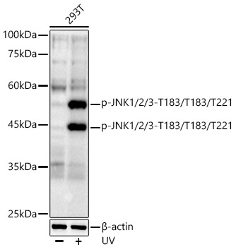
Western blot analysis of lysates from 293T cells, using Phospho-JNK1/2/3-T183/T183/T221 Rabbit mAb (A23099) at 1:1000 dilution. 293T cells were treated by UV at room temperature for 15-30 minutes.Secondary antibody: HRP-conjugated Goat anti-Rabbit IgG (H+L) (AS014) at 1:10000 dilution.Lysates/proteins: 25μg per lane.Blocking buffer: 3% nonfat dry milk in TBST.Detection: ECL Basic Kit (RM00020).Exposure time: 90s.
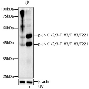
Western blot analysis of lysates from C6 cells, using Phospho-JNK1/2/3-T183/T183/T221 Rabbit mAb (A23099) at 1:1000 dilution. C6 cells were treated by UV at room temperature for 15-30 minutes.Secondary antibody: HRP-conjugated Goat anti-Rabbit IgG (H+L) (AS014) at 1:10000 dilution.Lysates/proteins: 25μg per lane.Blocking buffer: 3% nonfat dry milk in TBST.Detection: ECL Basic Kit (RM00020).Exposure time: 90s.
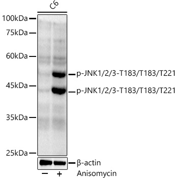
Western blot analysis of lysates from C6 cells, using Phospho-JNK1/2/3-T183/T183/T221 Rabbit mAb (A23099) at 1:1000 dilution. C6 cells were treated by Anisomycin (25 μg/mL) at 37℃ for 20 minutes.Secondary antibody: HRP-conjugated Goat anti-Rabbit IgG (H+L) (AS014) at 1:10000 dilution.Lysates/proteins: 25μg per lane.Blocking buffer: 3% nonfat dry milk in TBST.Detection: ECL Basic Kit (RM00020).Exposure time: 180s.
Immunohistochemistry analysis of paraffin-embedded Human colon carcinoma using Phospho-JNK1/2/3-T183/T183/T221 Rabbit mAb (AP0631) at dilution of 1:200 (40x lens). High pressure antigen retrieval performed with 0.01M Citrate Bufferr (pH 6.0) prior to IHC staining.
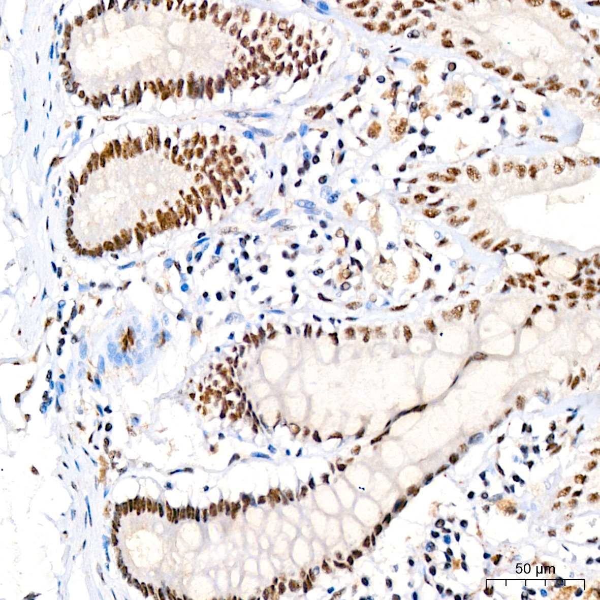
Immunohistochemistry analysis of paraffin-embedded Human colon using Phospho-JNK1/2/3-T183/T183/T221 Rabbit mAb (AP0631) at dilution of 1:200 (40x lens). High pressure antigen retrieval performed with 0.01M Citrate Bufferr (pH 6.0) prior to IHC staining.
Immunohistochemistry analysis of paraffin-embedded Human lung squamous carcinoma tissue using Phospho-JNK1/2/3-T183/T183/T221 Rabbit mAb (AP0631) at dilution of 1:200 (40x lens). High pressure antigen retrieval performed with 0.01M Citrate Bufferr (pH 6.0) prior to IHC staining.

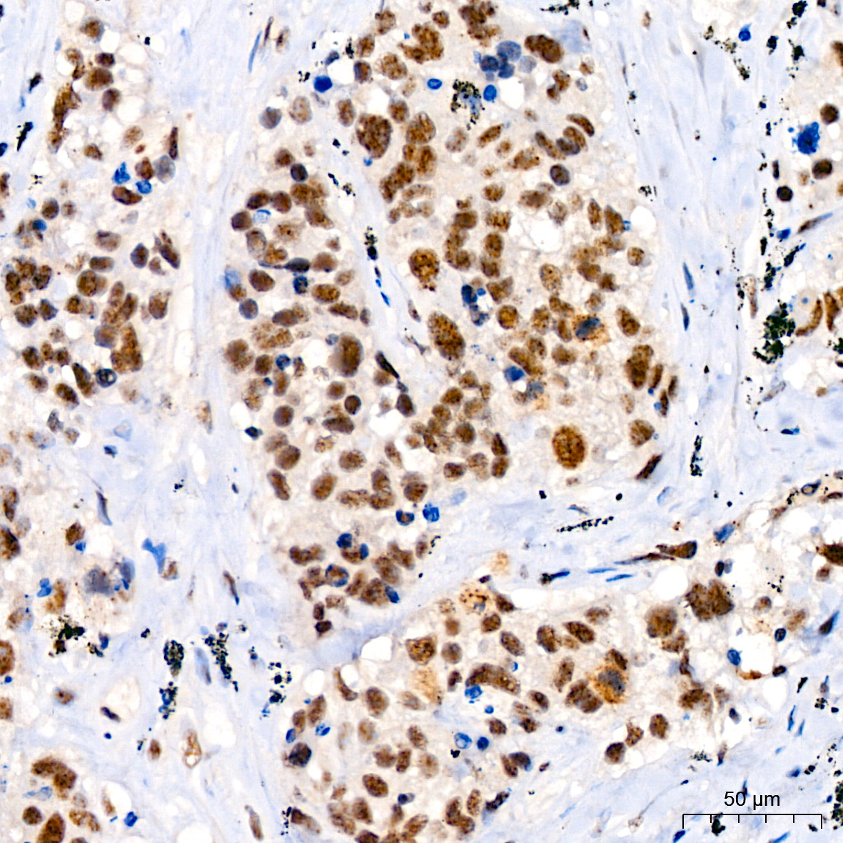
Immunohistochemistry analysis of paraffin-embedded Human tonsil using Phospho-JNK1/2/3-T183/T183/T221 Rabbit mAb (AP0631) at dilution of 1:200 (40x lens). High pressure antigen retrieval performed with 0.01M Citrate Bufferr (pH 6.0) prior to IHC staining.
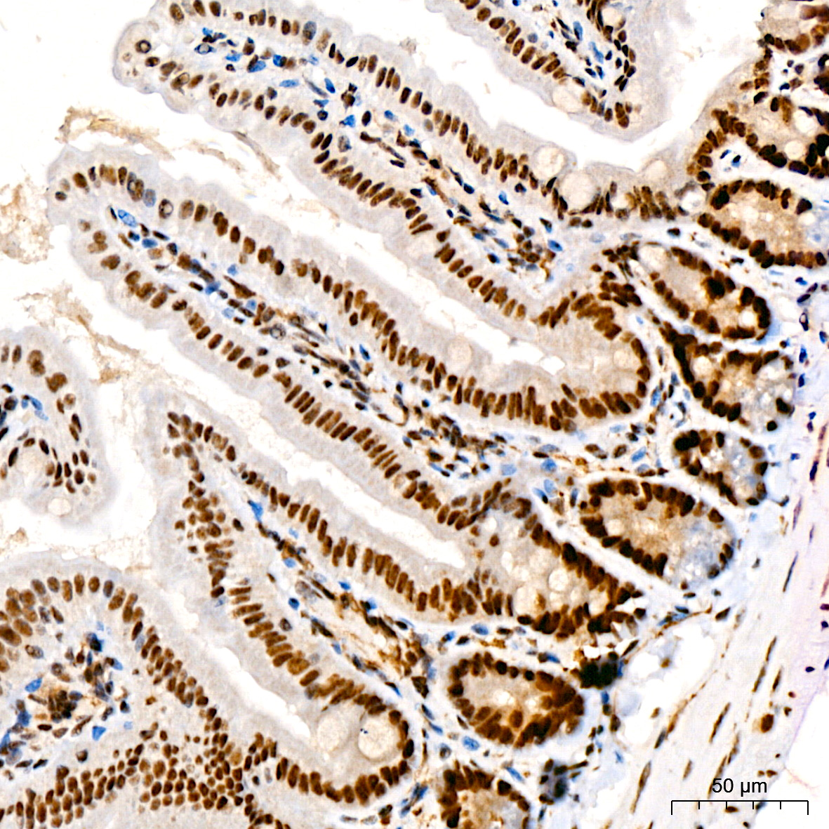
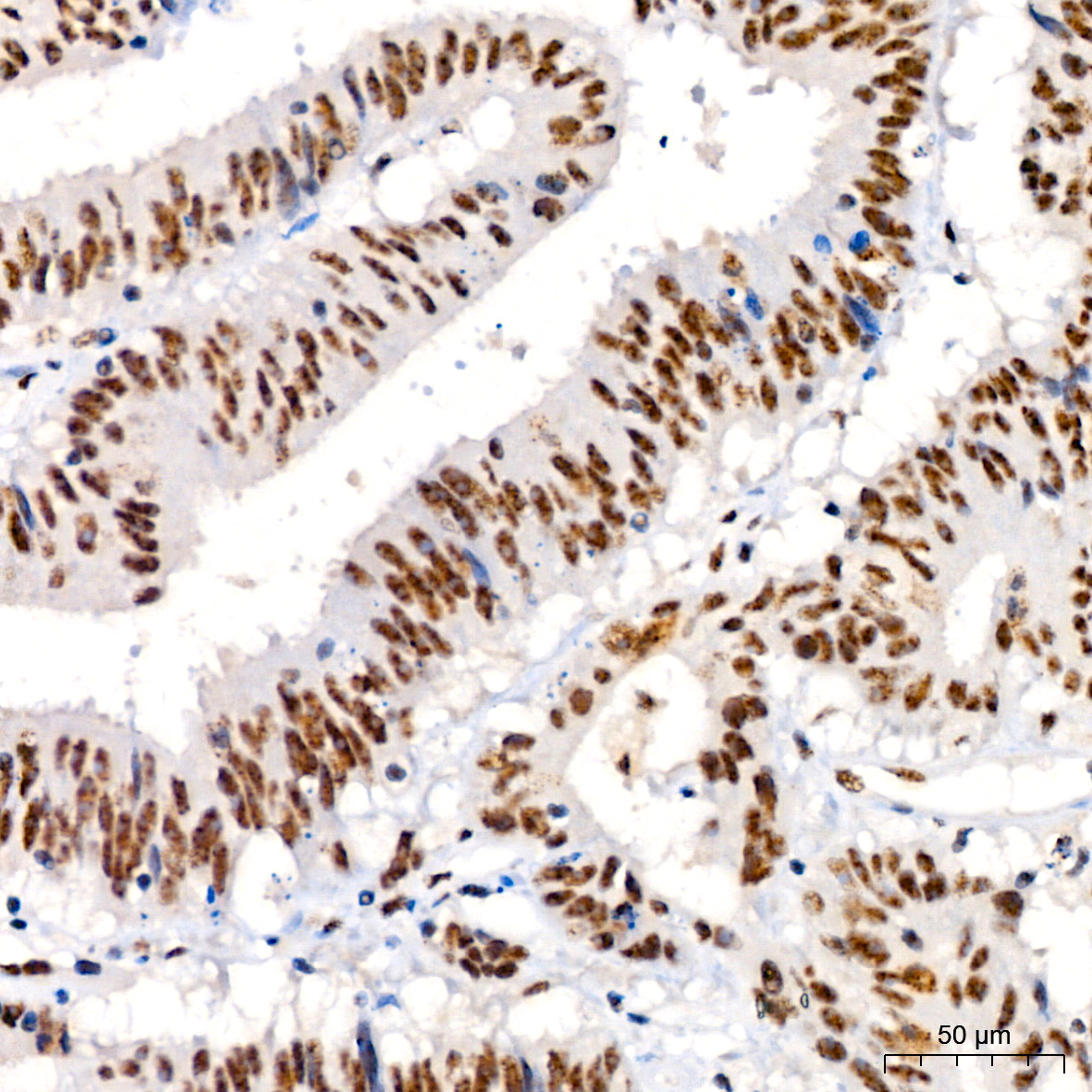
Immunohistochemistry analysis of paraffin-embedded Mouse colon using Phospho-JNK1/2/3-T183/T183/T221 Rabbit mAb (AP0631) at dilution of 1:200 (40x lens). High pressure antigen retrieval performed with 0.01M Citrate Bufferr (pH 6.0) prior to IHC staining.
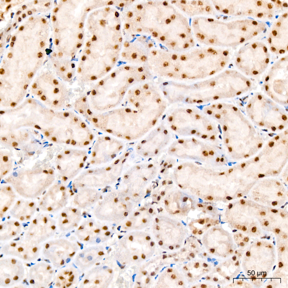
Immunohistochemistry analysis of paraffin-embedded Rat kidney using Phospho-JNK1/2/3-T183/T183/T221 Rabbit mAb (AP0631) at dilution of 1:200 (40x lens). High pressure antigen retrieval performed with 0.01M Citrate Bufferr (pH 6.0) prior to IHC staining.
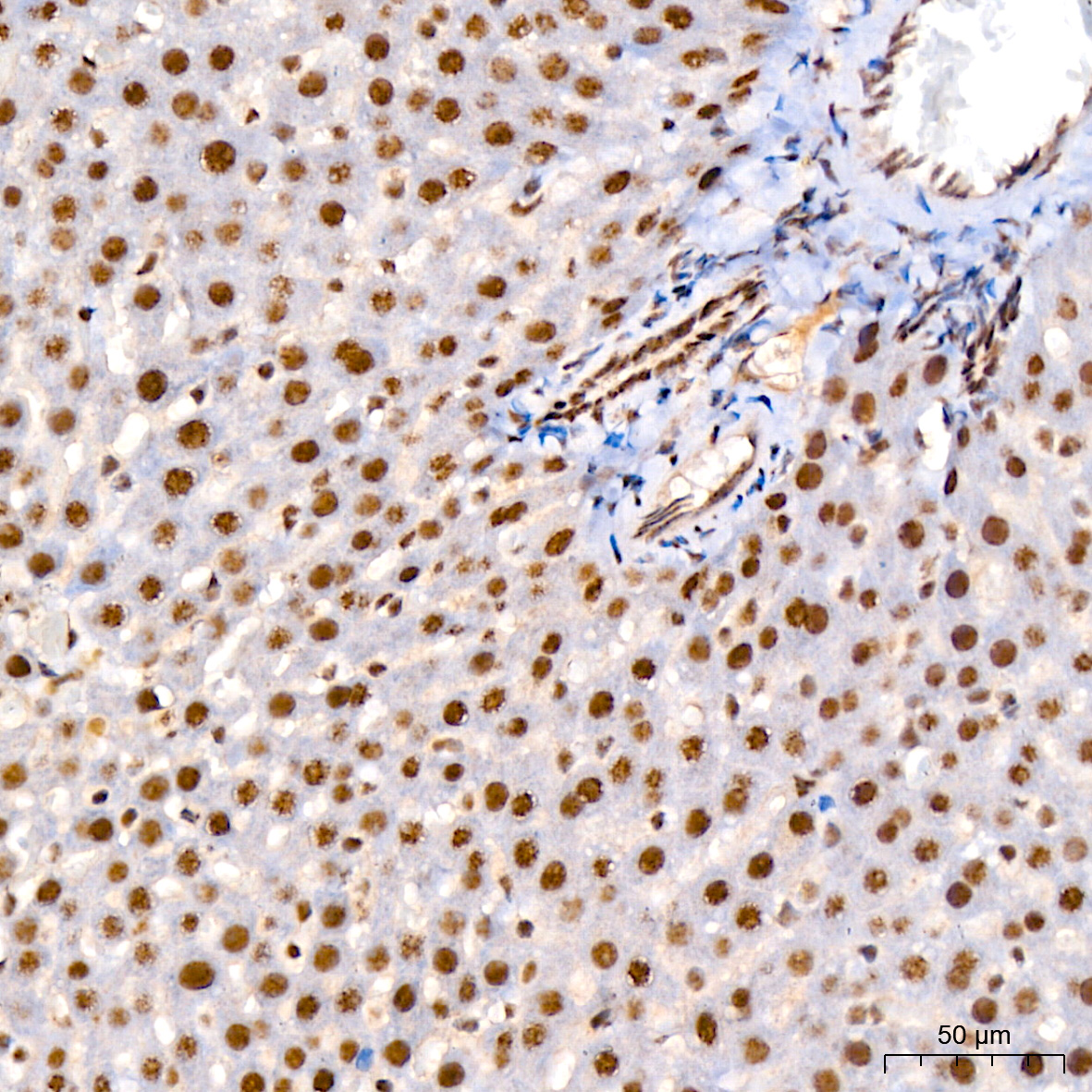
Immunohistochemistry analysis of paraffin-embedded Rat liver using Phospho-JNK1/2/3-T183/T183/T221 Rabbit mAb (AP0631) at dilution of 1:200 (40x lens). High pressure antigen retrieval performed with 0.01M Citrate Bufferr (pH 6.0) prior to IHC staining.
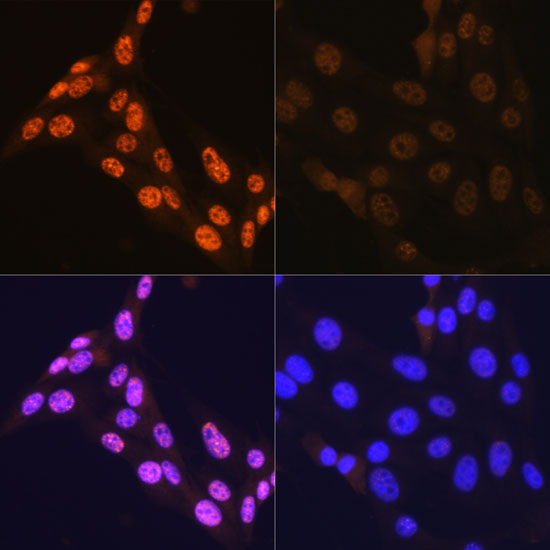
Immunofluorescence analysis of NIH-3T3 cells using Phospho-JNK1/2/3-T183/T183/T221 Rabbit mAb (AP0631).NIH-3T3 cells were treated by Anisomycin (25 μg/mL) at 37℃ for 30 minutes after serum-starvation overnight. Secondary antibody: Cy3-conjugated Goat anti-Rabbit IgG (H+L) (AS007) at 1:500 dilution. Blue: DAPI for nuclear staining.
Publishing research using AP0631? Please let us know so that we can cite the reference in this datasheet.
请输入产品标签上的lot号,例如4000000001
The protein encoded by this gene is a member of the MAP kinase family. MAP kinases act as an integration point for multiple biochemical signals, and are involved in a wide variety of cellular processes such as proliferation, differentiation, transcription regulation and development. This kinase is activated by various cell stimuli, and targets specific transcription factors, and thus mediates immediate-early gene expression in response to cell stimuli. The activation of this kinase by tumor-necrosis factor alpha (TNF-alpha) is found to be required for TNF-alpha induced apoptosis. This kinase is also involved in UV radiation induced apoptosis, which is thought to be related to cytochrom c-mediated cell death pathway. Studies of the mouse counterpart of this gene suggested that this kinase play a key role in T cell proliferation, apoptosis and differentiation. Several alternatively spliced transcript variants encoding distinct isoforms have been reported. [provided by RefSeq, Apr 2016]
* 该数据来源于赛特新思(citexs.com)
来自于发表文献,仅供参考所用,ABclonal不承担任何责任。
暂无以MAPK8/MAPK9/MAPK10为关键词搜索的文献记录。
该服务由赛特新思提供,上述关联分析来自已发表文献,仅供参考所用,ABclonal不承担任何责任。
首先,一般抗体不推荐客户回收利用,抗体使用之后缓冲体系已经发生改变,不同客户在回收抗体的保存条件上也会有差异,所以抗体回收使用效果无法保证。另外,ABclonal公司也做过一批抗体回收验证测试,测试结果显示不同抗体可回收次数不同,一般效价越高的抗体,可重复使用的次数越多,客户可根据实验情况来确定。
注:我们将孵育完毕后剩余的抗体回收到离心管中置于4℃保存,效价高的抗体可至少保存1周,至少重复利用3次。
武汉爱博泰克生物(ABclonal)科技有限公司是国产品牌,她成立于2011年,公司依托ABclonal美国波士顿抗体与蛋白研发中心、中国光谷生物城(武汉)抗体生产基地以及上海张江分子酶研发中心,凝聚了十余位来自哈佛大学、麻省理工、复旦大学、上海交大、中科院生化细胞所和武汉大学的全球知名分子和免疫学方面博士,组成我们的科学家团队,通过聚焦抗体与酶核心技术,致力于打破国际技术的垄断,将公司打造成为科研工具和诊断原料的国内领导品牌,乃至弯道超越国际巨头。 我们拥有包括兔多克隆抗体、小鼠单克隆抗体、兔单克隆抗体的生产研发平台,同时也有包括WB,IHC,IF,IP,CHIP在内的检测平台,我们对每一支自产的抗体进行了严格的检测。当然,我们部分直销地区也可以帮客户代购进口品牌的产品。同时也有抗体定制服务。ABclonal抗体优势:1,严自检,保质量;2产品多,指标全;3,价格低,货期短。注:ABclonal抗体价格体系详情见附件
ABclonal抗体成分在平时工作当中,常会有客户咨询我们的抗体用的什么buffer进行保存,一般来说,我们的buffer的成分是:PBS含0.03%的proclin300、0.05%牛血清白蛋白、50%甘油;也有一些是PBS含0.03%的proclin300,50%甘油。防腐剂 Proclin 300活性成分主要是2-甲基-4-异噻唑啉-3-酮(MCI)和5-氯-2-甲基-4-异噻唑啉-3-酮(CMCI)。ProClin生物灭活剂能够迅速穿透细胞膜,抑制对细胞呼吸至关重要的特定酶,因此一接触微生物有机体就会立即抑制细胞活性。ProClin的多个特定毒性位点可以防止微生物产生高水平的耐药性。

i_FC_01.jpg?t=1701849808)